Native T1 is independently predictive of aerobic functional capacity by VO2max in long-term follow-up of individuals with mild initial COVID disease
European Heart Journal


Abstract
Cardiovascular inflammatory involvement is an increasingly recognized sequela of COVID-19 disease. Cardiac magnetic resonance imaging (CMR) studies recorded subtle myocardial inflammatory changes, non-ischemic myocardial late gadolinium enhancement and pericarditis, also in previously well individuals with a mild initial course of disease.
To investigate the long-term cardiovascular consequences of COVID-19 on exercise intolerance and other symptoms using advanced CMR imaging and cardiopulmonary exercise testing (CPET).
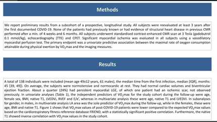
We report preliminary results from a subcohort of a prospective, longitudinal study. All subjects were reevaluated at least 3 years after the first documented COVID-19. None of the patients had previously known or had evidence of structural heart disease in previous CMR performed after a minimum of 4 weeks and 6 months. All subjects underwent standardised contrast-enhanced CMR scan at 3 Tesla (gadobutrol 0.1 mmol/kg), echocardiography and CPET. Significant myocardial ischemia was evaluated in all subjects using a vasodilatory myocardial perfusion test. The primary endpoint was a univariate predictive association between the maximal rate of oxygen consumption attainable during physical exertion by VO2max and the imaging measures.
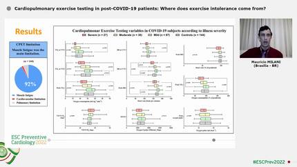
A total of 138 individuals were included (mean age 49±12 years, 61 males), the median time from the first infection, median [IQR], months: 45 [39, 49]). On average, the subjects were normotensive and normocardic at rest. They had normal cardiac volumes and biventricular ejection fraction. About a quarter (24%) had persistent myocardial LGE, of which one patient had an ischemic scar, not observed previously. In univariate analyses (Table 1), the independent predictors of VO2max for the study cohort during the follow-up were age, female sex, BMI, native T1, LVEDVi, RVEF and E/e’, whereas in multivariate analysis these were age, native T1 and LVEDVi. In subanalyses for gender, in males, in multivariate analysis LA area was the sole predictor of VO2max during the follow-up, while in the females, these were age, BMI and native T1. Figure 1 shows that VO2max values of post-COVID-19 patients were lower compared to the expected VO2max values based on the cardiorespiratory fitness reference database FRIEND, with a statistically significant positive correlation. Furthermore, the native T1 showed inverse correlation with VO2max values in the study cohort.
In individuals without previously known structural heart disease and mild initial COVID-19 disease, age, female sex, BMI, LVEDVi, RVEF, E/e', and native T1 were univariate predictors of VO2max at long-term follow-up. In multivariate analysis age, LVEDVi and native T1 were independent predictors of VO2max at follow-up. Patients with a history of COVID-19 achieved lower VO2max values compared to the reference values measured in the FRIEND registry. Native T1 showed an inverse correlation with VO2max values.
Contributors

M Ganbat
Author


E Nagel
Author
Institute for Experimental and Translational Cardiovascular Imaging Frankfurt , Germany
V Puntmann
Author
You may be interested in