Novel systematic processing of cardiac magnetic resonance imaging identifies target regions associated with infarct-related ventricular tachycardia

EP Europace Journal

19 September 2024
Organised by: Logo
ESC Journals ARRHYTHMIAS AND DEVICE THERAPY IMAGING Cardiac Magnetic Resonance (CMR) BASIC SCIENCE

Abstract

AbstractAims

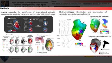
There is lack of agreement on late gadolinium enhancement cardiac magnetic resonance (LGE-CMR) imaging processing for guiding ventricular tachycardia (VT) ablation. We aim at developing and validating a systematic processing approach on LGE-CMR images to identify VT corridors that contain critical VT isthmus sites.

Methods and results

This is a translational study including 18 pigs with established myocardial infarction and inducible VT undergoing in vivo characterization of the anatomical and functional myocardial substrate associated with VT maintenance. Clinical validation was conducted in a multicentre series of 33 patients with ischaemic cardiomyopathy undergoing VT ablation. Three-dimensional LGE-CMR images were processed using systematic scanning of 15 signal intensity (SI) cut-off ranges to obtain surface visualization of all potential VT corridors. Analysis and comparisons of imaging and electrophysiological data were performed in individuals with full electrophysiological characterization of the isthmus sites of at least one VT morphology. In both the experimental pig model and patients undergoing VT ablation, all the electrophysiologically defined isthmus sites (n = 11 and n = 19, respectively) showed overlapping regions with CMR-based potential VT corridors. Such imaging-based VT corridors were less specific than electrophysiologically guided ablation lesions at critical isthmus sites. However, an optimized strategy using the 7 most relevant SI cut-off ranges among patients showed an increase in specificity compared to using 15 SI cut-off ranges (70 vs. 62%, respectively), without diminishing the capability to detect VT isthmus sites (sensitivity 100%).

Conclusion

Systematic imaging processing of LGE-CMR sequences using several SI cut-off ranges may improve and standardize procedure planning to identify VT isthmus sites.

Contributors

Alba Ramos-Prada
Alba Ramos-Prada

Author

Spanish National Centre for Cardiovascular Research Madrid , Spain

Andrés Redondo-Rodríguez

Author

Spanish National Centre for Cardiovascular Research Madrid , Spain

Andreu Porta-Sánchez

Author

Hospital Clinic of Barcelona Barcelona , Spain

Rachel M A ter Bekke

Author

Cardiovascular Research Institute Maastricht (CARIM) Maastricht , Netherlands (The)

Jose Luis Merino
Jose Luis Merino

Author

La Paz University Hospital Madrid , Spain

Matthijs Cluitmans
Matthijs Cluitmans

Author

Cardiovascular Research Institute Maastricht (CARIM) Maastricht , Netherlands (The)

Robert J Holtackers
Robert J Holtackers

Author

Maastricht University Medical Centre (MUMC) Maastricht , Netherlands (The)

Gonzalo Pizarro
Gonzalo Pizarro

Author

National Centre for Cardiovascular Research (CNIC) Madrid , Spain

Borja Ibáñez
Borja Ibáñez

Author

National Centre for Cardiovascular Research CNIC AND Fundacion Jimenez Diaz Hospital Madrid , Spain

David Calvo
David Calvo

Author

San Carlos Clinical Hospital Madrid , Spain

David Filgueiras-Rama

Author

Spanish National Centre for Cardiovascular Research Madrid , Spain