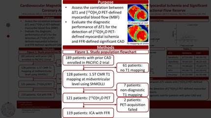
Functional stress imaging to predict abnormal coronary fractional flow reserve: the PACIFIC 2 study
European Heart Journal


Abstract
The diagnostic performance of non-invasive imaging in patients with prior coronary artery disease (CAD) has not been tested in prospective head-to-head comparative studies. The aim of this study was to compare the diagnostic performance of qualitative single-photon emission computed tomography (SPECT), quantitative positron emission tomography (PET), and qualitative magnetic resonance imaging (MRI) in patients with a prior myocardial infarction (MI) or percutaneous coronary intervention (PCI).
In this prospective clinical study, all patients with prior MI and/or PCI and new symptoms of ischaemic CAD underwent 99mTc-tetrofosmin SPECT, [15O]H2O PET, and MRI, followed by invasive coronary angiography with fractional flow reserve (FFR) in all coronary arteries. All modalities were interpreted by core laboratories. Haemodynamically significant CAD was defined by at least one coronary artery with an FFR ≤0.80. Among the 189 enrolled patients, 63% had significant CAD. Sensitivity was 67% (95% confidence interval 58–76%) for SPECT, 81% (72–87%) for PET, and 66% (56–75%) for MRI. Specificity was 61% (48–72%) for SPECT, 65% (53–76%) for PET, and 62% (49–74%) for MRI. Sensitivity of PET was higher than SPECT (
In this prospective head-to-head comparative study, SPECT, PET, and MRI did not show a significantly different accuracy for diagnosing FFR defined significant CAD in patients with prior PCI and/or MI. Overall diagnostic performances, however, were discouraging and the additive value of non-invasive imaging in this high-risk population is questionable.
Contributors

Roel S Driessen
Author
Pepijn A van Diemen
Author
Pieter G Raijmakers
Author
Juhani Knuuti
Author


S Richard Underwood
Author
Imperial College London London , United Kingdom of Great Britain & Northern Ireland


Lourens F H J Robbers
Author
Amsterdam University Medical Centre (AUMC) Amsterdam , Netherlands (The)

Martin B von Bartheld
Author
Peter M van de Ven
Author
Leonard Hofstra
Author
G Aernout Somsen
Author
Igor I Tulevski
Author
Ronald Boellaard
Author
Albert C van Rossum
Author
Ibrahim Danad
Author
Paul Knaapen
Author
You may be interested in