68 year-old female patient with exertional dyspnoea


The case
A 68-year-old female patient (BMI 20.8 kg/m2) complained of exertional dyspnoea:
- She had a history of composite graft replacement of the aortic root because of a bicuspid aortic valve with a dilated annulus 9 months ago.
- A DDD pacemaker was implanted due to postoperative atrioventricular (AV) block.
- A cardiopulmonary exercise test was performed on a treadmill using the modified Bruce protocol.
SPIROMETRY
9 PANEL PLOT OF THE CARDIOPULMONARY EXERCISE TEST
KEY PANELS
EXERCISE ECG
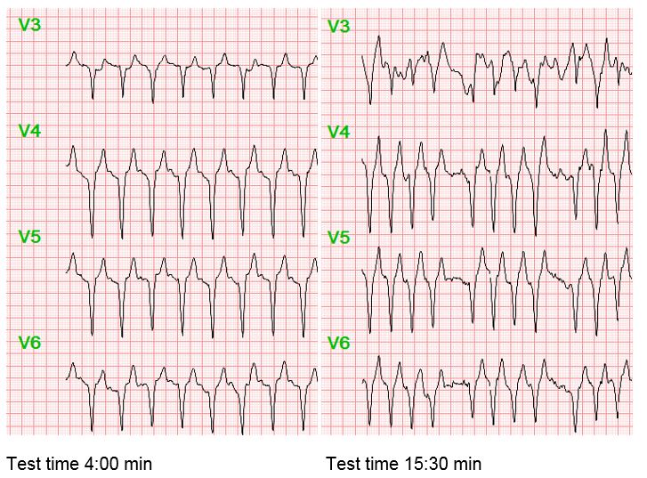
Test findings
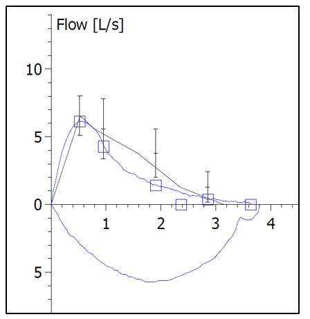
The resting spirometry showed a FVC of 3.6 l (116% predicted), a FEV1 of 2.4 l (89% predicted), and a FEV1/FVC of 65%. Estimated maximum voluntary ventilation (MVV, FEV1*40) was 96 l.
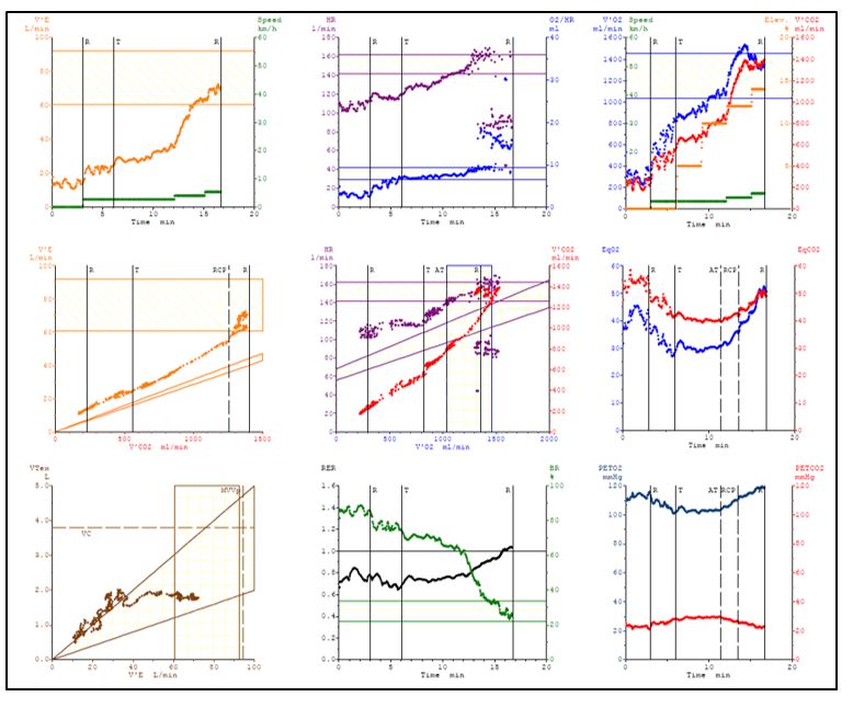
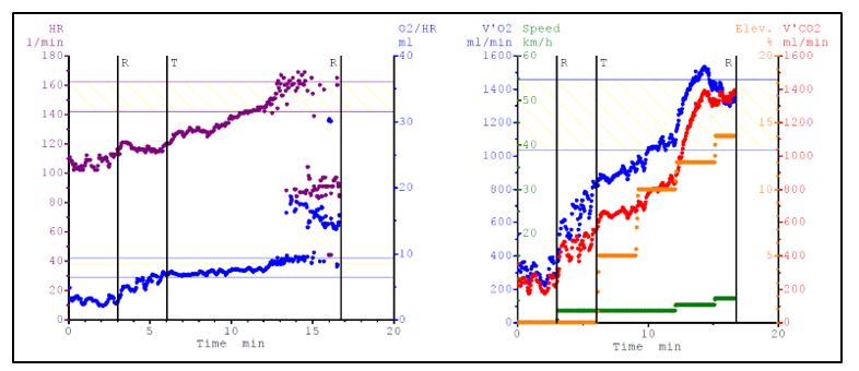
Cardiorespiratory fitness was normal with a peak VO2 of 20.5 ml/min/kg (5.9 MET, 106 % predicted) (Panel 3).
Blood pressure increased from 130/90 mmHg to 160/105 mmHg at peak exercise. Maximum respiratory exchange ratio was 1.04 (Panel 8). The patient was exhausted at the end of the test (Borg 18/20) and complained of exertional dyspnoea.
Maximum ventilation was 69 l (Panel 1). Breathing reserve ((1-VEmax/MVV)*100) was 27% (Panel 8). Respiratory efficiency, determined by the VE/VCO2 slope was 38 (Panel 4). Peak PETCO2 was 29 mmHg (Panel 9). The O2 pulse (VO2/heart rate) increased to 15.8 ml (199% of predicted, Panel 2). At the end of the test, a sudden drop in oxygen uptake (Panel 3) was detected.
You may be interested in