
References
Question 1:
1. Sharma S, Drezner JA, Baggish A, et al. International recommendations for electrocardiographic interpretation in athletes. Eur Heart J 2018; 39: 1466–1480.
2. Malhotra A, Dhutia H, Gati S, et al. Anterior T-Wave Inversion in Young White Athletes and Nonathletes: Prevalence and Significance. J Am Coll Cardiol. 2017;69(1):1-9.
Question 2:
1. Caudron J, Fares J, Vivier PH, et al. Diagnostic accuracy and variability of three semi-quantitative methods for assessing right ventricular systolic function from cardiac MRI in patients with acquired heart disease. Eur Radiol 2011; 21: 2111.
2. Galea N, Carbone I, Cannata D, et al. Right ventricular cardiovascular magnetic resonance imaging: normal anatomy and spectrum of pathological findings. Insights Imaging 2013; 4: 213.
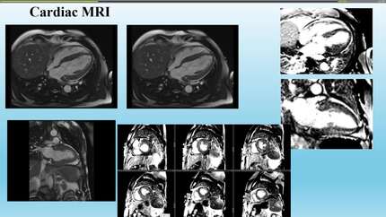
3. Licordari R, Trimarchi G, Teresi L, et al. Cardiac Magnetic Resonance in HCM Phenocopies: From Diagnosis to Risk Stratification and Therapeutic Management. J Clin Med 2023; 12: 12.
4. Dubrcy SW, Falk RH. Amyloid heart disease. Br J Hosp Med 2010; 71: 76–82.
5. Nemshah Y, Clavijo A, Sharma G. Amyloid heart disease. US Cardiol Rev 2018; 12: 113–118.
6. Hoigné P, Attenhofer Jost CH, Duru F, et al. Simple criteria for differentiation of Fabry disease from amyloid heart disease and other causes of left ventricular hypertrophy. Int J Cardiol 2006; 111: 413–422.
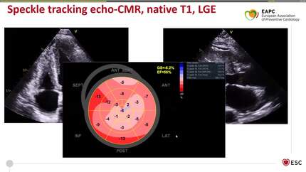
7. Lavall D, Vosshage NH, Geßner R, et al. Native T1 mapping for the diagnosis of cardiac amyloidosis in patients with left ventricular hypertrophy. Clin Res Cardiol 2023; 112: 334–342.
Question 3:
1. National Institute for Health and Care Excellence (NICE). Myeloma: diagnosis and management (NICE guideline NG35). Published 10 Feb 2016. Last updated 25 Oct 2018. Available at: https://www.nice.org.uk/guidance/ng35/.
2. Rajkumar SV. Updated Diagnostic Criteria and Staging System for Multiple Myeloma. Am Soc Clin Oncol Educ B 2016; e418–e423.
3. Kim MA, Kim CH, Oh BH, et al. Cardiac Amyloidosis Diagnosed by Endomyocardial Biopsy. Korean J Intern Med 1988; 3: 148.
4. Granstam SO, Rosengren S, Vedin O, et al. Evaluation of patients with cardiac amyloidosis using echocardiography, ECG and right heart catheterization. Amyloid 2013; 20: 27–33.
5. Paeng JC, Choi JY. Nuclear Imaging for Cardiac Amyloidosis: Bone Scan, SPECT/CT, and Amyloid-Targeting PET. Nucl Med Mol Imaging (2010) 2021; 55: 61.
6. Phull P, Sanchorawala V, Connors LH, et al. Monoclonal gammopathy of undetermined significance in systemic transthyretin amyloidosis (ATTR). Amyloid 2018; 25: 62.
Notes to editor
Authors information:
- Dr Emmanouil Androulakis,
- Dr Szymon Musiol, Royal United Hospitals Bath NHS Foundation Trust
- Dr Michael Papadakis, St George’s University Hospitals NHS Foundation Trust
- Dr Maria Teresa Tome Esteban, St George’s University Hospitals NHS Foundation Trust