Zero-fluoroscopy, intracardiac echocardiography–only patent foramen ovale closure in early pregnancy: a case report
European Heart Journal - Case Reports


Abstract
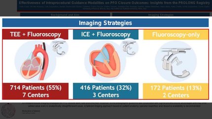
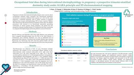
Transcatheter closure of a patent foramen ovale (PFO) is an established strategy for secondary prevention of cryptogenic stroke in selected patients. During pregnancy, the timing and method of closure require individualized assessment of recurrent embolic risk and maternal–fetal procedural risk. In selected first-trimester patients, avoiding fluoroscopy, contrast, procedural transoesophageal echocardiography (TEE), and sedation may be desirable when adequate alternative imaging is available.
A 34-year-old woman at 7 weeks’ gestation presented with acute ischaemic stroke. Work-up demonstrated a non-lacunar cortical infarct, long-tunnel PFO with a large provoked right-to-left shunt, and no alternative embolic source; the Risk of Paradoxical Embolism score was 9. After multidisciplinary discussion, early percutaneous closure was performed using a completely zero-fluoroscopy, intracardiac echocardiography (ICE)-only workflow under local anaesthesia, without procedural TEE. Key adaptations included use of a pre-shaped transseptal sheath for stable cannulation and a left atrial rail using a pre-shaped support wire. Stop-flow balloon sizing was performed and agitated saline was injected through the delivery sheath as an adjunctive visual check of left atrial position prior to device release. A 25/18-mm Amplatzer Talisman PFO occluder was deployed successfully with no complications.
This case demonstrated the feasibility of zero-fluoroscopy, zero-contrast PFO closure under ICE-only guidance in a carefully selected first-trimester pregnant patient. The combination of stable sheath engagement, a pre-shaped left atrial support-wire rail, stop-flow balloon sizing, and sheath-through agitated saline injection facilitated controlled device deployment without fluoroscopy or contrast. This approach depends on operator expertise and institutional familiarity with ICE-guided structural intervention.
Contributors

SungHo Jo
Author
Tae-Hoon Kim
Author
Yong-Joon Lee
Author
KyeongHo Yun
Author

Lina Ya'qoub
Author
A Ashika
Author
Deepti Ranganathan
Author
You may be interested in