Computed tomography pitfalls and diagnostic value in a patient with post-infarction ventricular septal defect, cardiogenic shock, and iatrogenic type B aortic dissection during extracorporeal membrane oxygenation support: a case report
European Heart Journal - Case Reports


Abstract
Mechanical complications after acute myocardial infarction are associated with high mortality. When a post-infarction ventricular septal defect is complicated by cardiogenic shock and acute aortic dissection during escalation of mechanical circulatory support, multimodality imaging becomes critical for diagnosis and management.
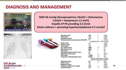
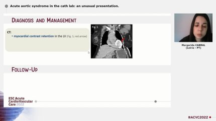
A 56-year-old man presented with inferior ST-segment elevation myocardial infarction and underwent urgent coronary angiography, which showed proximal right coronary artery occlusion with only partial reperfusion. Transthoracic echocardiography identified a large inferior ventricular septal defect with a holosystolic left-to-right shunt. Because of haemodynamic deterioration and high surgical bleeding risk after dual antiplatelet therapy, an intra-aortic balloon pump was inserted, followed by femoro-femoral extracorporeal membrane oxygenation for refractory cardiogenic shock. Computed tomography demonstrated type B aortic dissection with partial false lumen thrombosis and showed that the intra-aortic balloon pump had been inadvertently positioned within the false lumen, prompting immediate removal. Computed tomography also revealed extracorporeal membrane oxygenation-related enhancement artefacts. Follow-up computed tomography showed no clear progression. On Day 5, the patient developed refractory ventricular fibrillation. Transoesophageal echocardiography demonstrated severe aortic root and proximal ascending aortic thrombosis, absent aortic valve opening, and minimal residual perfusion. The patient died shortly afterwards.
This case highlights the diagnostic and management value of advanced computed tomography in a patient with post-infarction ventricular septal defect, cardiogenic shock, and iatrogenic type B aortic dissection during extracorporeal membrane oxygenation support. It also illustrates important computed tomography pitfalls related to competing blood streams and watershed artefacts under peripheral extracorporeal membrane oxygenation.
Contributors

Adrià Ruiz-López
Author
Mario Salido Iniesta
Author
Sònia Gómez Revelles
Author
Claudia Taroncher Domingo
Author
David Viladés Medel
Author
Constantin Kühl
Author
Arif Albulushi
Author
Ahsan Aftab Khan
Author
Deepti Ranganathan
Author
You may be interested in