Case series: the right parasternal view and its prognostic value in detecting pathological abnormalities on transthoracic echocardiography
European Heart Journal - Case Reports


Abstract
Two-dimensional echocardiography is the most widely used imaging modality for immediate diagnosis, due to its easy access and cost-effectiveness. International guidelines recommend four standard views to perform a comprehensive minimum dataset for every adult patient, namely left parasternal, apical, subcostal, and suprasternal views.
In this case series, we revisit the right parasternal view (RPV). Although it is infrequently used in routine clinical practice, it provides valuable complementary information, particularly when abnormalities are found on the minimum dataset or when measurement results are inconsistent. Including this view in the imaging protocol adds only a few extra minutes but can yield important additional diagnostic information.
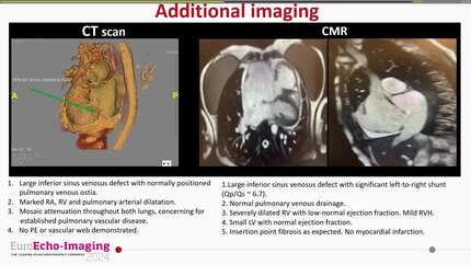
Presented are four cases in which the complementary use of RPV provided decisive diagnostic information that was either missed or underestimated by standard echocardiographic windows and influenced clinical management, re-emphasizing the value of adding this view during a routine examination.
Contributors

Neve Moore
Author
Alyssa Lacerna
Author
Juan Ortegarestrepo
Author
Roxy Senior
Author
Rajdeep Khattar
Author
Wei Li
Author
Sitali Mushemi-Blake
Author
Rita Pavasini
Author
Alejandro Ismael Cordero
Author
Ivan Wong
Author
Malak Benabdellah
Author
Deepti Ranganathan
Author
You may be interested in