Case reports of a double threat: when cancer and hidden congenital heart defects collide: malignant tamponade unveils a hidden pulmonary venous anomaly
European Heart Journal - Case Reports


Abstract
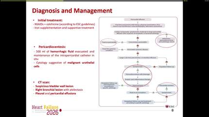
Malignant pericardial effusion is a life-threatening manifestation of advanced non-small-cell lung cancer (NSCLC). Cardiac tamponade as an initial presentation is uncommon, and coexistent pathologies can complicate diagnosis and management.
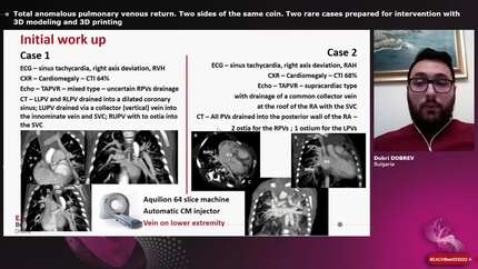
A 72-year-old man with mild chronic obstructive pulmonary disease (COPD) and a remote 2-pack-year smoking history was admitted with COVID-19 infection and possible superimposed pneumonia. Initial imaging revealed pericardial inflammation and a small effusion, raising concern for pericarditis. Subsequent echocardiography demonstrated a large pericardial effusion with tamponade physiology requiring pericardiocentesis, while cardiac magnetic resonance and computed tomography suggested possible malignancy. Cytology from pericardial and pleural fluid confirmed adenocarcinoma and bronchoscopy established stage IV epidermal growth factor–mutant NSCLC with cerebellar metastases. The patient experienced recurrent malignant effusions, managed successfully with balloon pericardiotomy and was treated with Osimertinib, resulting in sustained remission of effusions and clinical recovery. Careful review of multimodality imaging also revealed partial anomalous pulmonary venous return with significant left-to-right shunting, which may have exerted a protective hemodynamic effect on tamponade physiology. This may suggest a potential, though speculative, contribution of the anomalous pulmonary venous return to the observed tamponade physiology. At 2-year follow-up, the patient remains clinically and hemodynamically stable.
This case illustrates the interesting duality of how multiple concurrent pathologies—including COVID-19 pneumonia, pericarditis, malignant pleuro-pericardial effusions, metastatic lung adenocarcinoma, and PAPVR—can coexist and probably shape the clinical course. The case underscores the indispensable role of multimodality imaging, molecular profiling, and minimally invasive pericardial interventions in guiding diagnosis, therapy, and prognosis in complex cardio-oncology presentations.
Contributors

Alexis Phyllip Rodriguez
Author

Sarandeep Kaur Marwaha
Author
Alessandro Palmieri
Author
Giulia Bruno
Author
Deepti Ranganathan
Author
You may be interested in