The role of opa1 in mitochondrial remodeling and profibrotic activation of cardiac fibroblasts
Cardiovascular Research


Abstract
Cardiac fibroblasts (CFs) contribute to fibrosis by differentiating into myofibroblasts. Mitochondrial dynamics, regulated by fission/fusion, are essential for cell homeostasis. Optic Atrophy 1 (OPA1), a dynamin-related GTPase supporting inner mitochondrial membrane fusion, has cardioprotective functions, but its role in CFs is unclear.
To investigate mitochondrial dynamics in CFs and whether OPA1 modulation influences their differentiation upon TGFβ1 stimulation.
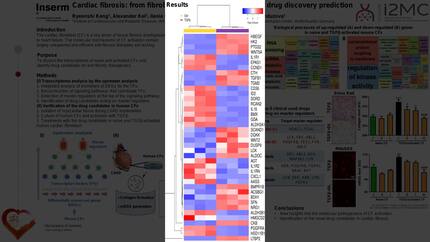
CFs from C57Bl6J mice were exposed to TGFβ1 stimulation and transfected with OPA1 siRNA. Mitochondrial morphology was evaluated by Mitotracker/MiNA, function by Seahorse OCR and Western blotting, and transcriptomic changes by RNA-seq/GO analysis. CF activation was assessed by αSMA and procollagen expression in 2D cultures and 3D spheroids.
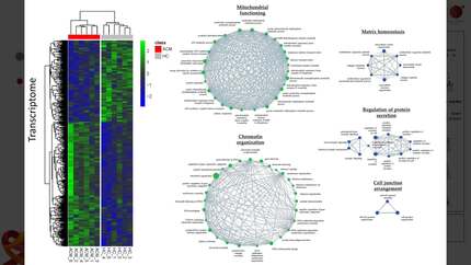
CFs exposed to TGFβ1 showed a dynamic remodeling of mitochondria: increased fusion at 16h (1.29-fold vs CTR a.u) followed by fragmentation at 24h (0.87-fold vs CTR a.u). Fluorescence intensity per cell increased (16h: 3.63-fold; 24h: 2.11-fold; 48h: 2.02-fold), reflecting enhanced activity. OPA1 expression was transiently reduced at 16h (0.82-fold), recovered at 24–48h, and significantly upregulated at 72h (1.32-fold, p<0.05, N=3). OPA1 silencing reduced gene (log2FC=-2.43, N=2) and protein levels (log2FC=-6.3, p<0.0001, N=3), increased fission (0.69-fold vs scramble, p<0.05, N=3), and impaired mitochondrial function (OCR 0.42-fold vs scramble). GO enrichment of siOPA1 DEGs (n=769, padj<0.05, N=3) revealed mitochondrial function, oxidative stress, and ATP metabolism (GO:0046034, GO:0006979, GO:0032981, padj<0.001, N=3). RNA-seq identified 968 DEGs in TGFβ1-treated CFs (padj<0.05, N=3), enriched in extracellular matrix (ECM) organization (GO:0030198, GO:0032956, GO:0032963, padj<0.005, N=3). OPA1 silencing blunted this transcriptional response, negatively regulating differentiation-related pathways (GO:0031345, GO:0050866, GO:0030574, padj<0.005, N=3). Coherently, the reduced TGFβ1-induced activation was demonstrated by αSMA expression in immunofluorescence (5.24-fold vs Scr, p<0.001; 0.48-fold vs TGFβ1 Scr, p<0.01, N=3) and Western blot (2.37-fold vs Scr, p<0.01; 0.57-fold vs TGFβ1 Scr, p<0.05, N=3). Western blot also confirmed reduced intracellular procollagen levels (4.34-fold vs Scr, p<0.001; 0.36-fold vs TGFβ1 Scr, p<0.01, N=3). OPA1 silencing increased spheroid number but reduced their size/compaction (Sphere: Scr 21±4.63; siOPA1: 41±4.34; p<0.01; N=5; Diameter: Scr 225.4±14.58; siOPA1: 183±11.92; p<0.05; N=5), indicating impaired cell interactions and diminished myofibroblast activation in 3D, as assessed by αSMA levels (2.85-fold vs Scr; 0.36-fold vs TGFβ1 Scr).
OPA1 emerges as a central mediator of TGFβ1-driven fibrotic activation: its silencing disrupts mitochondrial dynamics and function, thereby blunting TGFβ1-induced ECM production and fibroblast-to-myofibroblast differentiation.
Contributors


You may be interested in