Interventricular septum displacement and hemodynamic evaluation across heart failure spectrum during exercise performance
European Heart Journal


Abstract
Impairment of ventricular interdependence may determine the typical exertional dyspnea in heart failure (HF). The implications of the pathophysiological insights of biventricular interaction has been poorly understood over time. We hypothesized that a comprehensive analysis of the interventricular septum (IVS) adaptations during exercise performance in HF may unlock the related mechanisms to the limited O2 uptake and impaired cardiac reserve.
Study objectives were to study the pathophysiology of biventricular interaction during exercise in HF exploring how the IVS curvature changes during exercise may impact on exercise performance
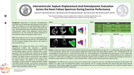
33 HF patients (20 HFpEF and 13 HFrEF) were prospectively enrolled, who underwent a combined cardiopulmonary exercise testing imaging (echoCPET) with RV 3D-imaging analysis and were compared with a control population. The RV chamber was assessed by 3D analysis and images were examined off-line using the 4D RV TomTec software. 3D septal curvature reconstruction was obtained by a 3D mesh of the mean curvature value, using a custom RV model software. The degree of IVS curvature was examined in 4 regions of the RV - inflow tract (RVIT), outflow tract (RVOT), apical, and body - and curvature measurements were acquired during end-diastole (ED) and end systole (ES) phases using a parametric curvature map.
An abnormal septal curve was found in patients with both HF phenotypes (HFpEF mean age 74.5 ± 7.5, 60% female; HFrEF mean age 64.6 ± 11.4, 21% female), typically with a more leftward configuration either at rest and during exercise (HFpEF: rest= −0.014±0.006 at ED, and −0.014±0.009 at ES; peak exercise= −0.011±0.01 at ED; HFrEF: rest: −0.011±0.008 at ED, −0.013±0.009 at ES; peak exercise= -0.012 ± 0.003 at ED, -0.012 ± 0.006 at ES) compared to controls (rest=−0.02±0.002 at ED, and −0.02±0.006 at ES; peak exercise −0.02±0.0.006 at ED, and −0.02±0.01 at ES, Figure 1). Furthermore, the degree of IVS curvature impairment showed a linear correlation with an impaired gas exchange performance as reflected in a lower peak VO2 in HF (HFpEF: r=0.54, p<0.001; HFrEF: r=0.60, p<0.001 at ED during exercise, Figure 2).
In HF, different IVS curvatures characterize HF patients when compared to controls, with differences related to HF phenotypes. The evidence of a right to left IVS displacement appears worth exploring, and it is strongly associated with reduced VO2 peak during exercise performance. These findings suggest the usefulness of evaluating how new and current therapeutic approaches may modify the negative septum displacement and overall cardiac hemodynamics.
Contributors



You may be interested in