Functional substrate identification using scar voltage mapping in ischemic fast ventricular tachycardia
European Heart Journal


Abstract
Mapping and ablating monomorphic fast ventricular tachycardias (FVT) with a cycle length <320 ms remains challenging due to hemodynamic instability. Identifying arrhythmogenic substrates during sinus rhythm (SR) is further complicated by rate-dependent functional properties of heterogeneous tissue (HT). Defining the functional boundaries of FVT isthmuses (FVTI) may enhance ablation outcomes.
We conducted an experimental study in 30 Large-White swine with anterior myocardial infarction. Cardiac magnetic resonance imaging (CMR) was performed for signal intensity (SI) mapping, followed by ultrahigh-density voltage mapping and electrophysiological studies at 4- and 16-weeks post-infarction. FVTIs were identified as corridors of high frequency electrograms spanning the electric diastole in reentrant propagation maps. Voltage heterogeneity maps during both FVT and SR (0.1-1.5 mV) were compared to assess the presence of functional substrates.
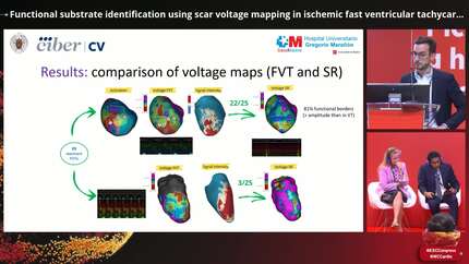
A total of 60 ventricular tachycardias were induced, with 27 classified as FVTs and mapped entirely. Among these, 25 demonstrated reentrant activation, allowing FVTI identification. All FVTI colocalized with SI-defined channels and voltage channels in FVT maps. Key findings from the voltage comparison between FVT and SR included: (1) an increased dense scar area (<0.1 mV) in FVT maps compared to SR (1.51 vs. 0.11 cm², p<0.001); (2) longer voltage channels sustaining FVTI during FVT compared to SR (18.7 vs. 14.6 mm, p=0.047); and (3) functional substrates detected in 81% of FVTIs, as evidenced by the presence of voltage channels bordered by dense scar in FVT maps, a feature observed in only four SR channels. Adjusting the lower voltage limit in SR voltage maps enabled retrospective identification of 88% of FVTIs.
Scar voltage heterogeneity mapping during FVT successfully identified all FVTI. The comparative analysis of voltage heterogeneity in FVT and SR allowed for precise delineation of functional isthmus borders, offering valuable insights into ventricular tachycardia substrate characterization that may lead the way to optimized ablation strategies.
Contributors




You may be interested in