Dynamics of R-wave amplitude, current of injury and impedance during lead advancement in patients undergoing left bundle branch area pacing monitored by intracardiac echocardiography
European Heart Journal


Abstract
Left bundle branch area pacing (LBBAP) is a widely used approach to physiological pacing. Proper lead placement is critical to avoid complications like septal perforation or loss of capture. Various electrical parameters (pacing impedance, current of injury (COI), R-wave amplitude) assess lead depth, but none are validated by imaging. Intracardiac echocardiography (ICE) enables real-time lead visualisation during deployment.
This study aims to evaluate electrical parameters at different lead depths using ICE and identify cut-off values used to diagnose perforation.
During LBBAP implantation, an ICE probe was introduced via the right femoral vein to visualise the septum, which for the purpose of the study was divided into three zones (Z1-Z3). Z1 covered 25% of septal thickness from the RV endocardium, Z2 the midseptum (25–75%), and Z3 the left ventricular septum, where conduction system capture (CSP) was confirmed at the final lead position. Electrical parameters—R-wave amplitude, COI at the lead tip (COItip) and ring (COIring) at 0.5-500 Hz filter settings during spontaneous rhythm, and pacing impedance were recorded at each zone. The COItip-to-COIring ratio (TRR) was calculated. CSP was assessed using accepted criteria.
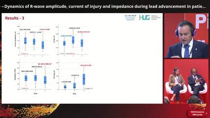
Forty-two patients (33 males, 78.6%, median age 70.5, IQR 63-76.5 years) were enrolled. Two participants were excluded due to unsuccessful lead placement while in the remainder, CSP was achieved in 39 (97.5%) after 2 (1-3) implantation attempts during the procedure. The results are shown in the figure. R-wave amplitude and pacing impedance showed significant reductions from Z2 to Z3 (P=0.001 and P=0.019, respectively). COItip showed a significant increase from Z1 to Z2 (P=0.031) and a decrease from Z2 to Z3 (P=0.038) to return to values which were comparable to those of Z1. The minimum value for COItip in Z3 at the final acceptable position was 5.2 mV. COIring increased significantly Z2 and Z3 (P=0.0001). TRR also significantly changed ( 9.62, IQR 4.42-17.23 vs. 11.80, IQR 3.94-23.21 vs. 2.02, IQR 1.27-10.76, P<0.0001) with a decrease from Z2 to Z3 (P=0.0001) . There were 4 cases (10%) of microperforation 3 of which showed lower spontaneous COI values than in Z3 (1.6, 2.88, 2.4 and 7.8 mV). No TRR values in Z3 were below 0.5 while only one of the four microperforation cases had values above this threshold.
ICE-guided LBBAP enables real-time monitoring of lead positioning. COI, impedance, and R-wave amplitude change significantly with lead advancement, particularly between Z2 and Z3. TRR<0.5 may help diagnose microperforation.
Contributors



You may be interested in