Brain lesions in patients with heart failure and atrial fibrillation
European Heart Journal


Abstract
Both patients with heart failure (HF) and patients with atrial fibrillation (AF) have an increased risk for MRI-detected brain lesions compared to patients without these diseases. Data on the interaction of AF and HF with regards to brain lesions are lacking.
We aimed to investigate the associations of HF and AF, and their interaction, with MRI-detected brain lesions in the Swiss-AF study.
We enrolled 3,418 patients into the multicenter Swiss-AF study across 14 centers in Switzerland. Patients were classified into four distinct groups based on their AF and HF status: patients without AF and HF (AF-/HF-), with HF but no AF (AF-/HF+), with AF but no HF (AF+/HF-), and with both AF and HF (AF+/HF+). Brain magnetic resonance imaging (bMRI) was performed to assess large non-cortical and cortical infarcts (LNCCI), small non-cortical infarcts (SNCI), white matter hyperintensities (WMH), and microbleeds (Mb). We used logistic regression models to examine the associations of AF and HF, and their interaction with each individual brain lesion type. At a second step, the same models were adjusted for age, sex, smoking, arterial hypertension, systolic blood pressure, diabetes mellitus, and a history of stroke or transient ischemic attack.
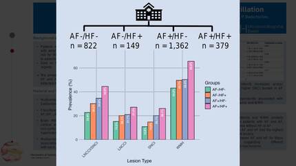
We included 2,712 patients with available bMRI. Mean age was 73 years and 30.8% were female. 822 (30.3%) patients were classified into the AF-/HF- group, 149 (5.5%) into the AF−/HF+ group, 1,362 (50.2%) into the AF+/HF− group, and 379 (14%) into AF+/HF+ group. Out of the 1,741 patients with AF, 90.1% were on anticoagulation and 17.6% on antiplatelet therapy compared to 9.1% and 58.7%, respectively, in patients without AF. Ischemic brain infarcts increased across the four groups with the highest prevalence in the AF+/HF+ group (Figure 1). This was driven by a higher burden of SNCI in the AF groups, while LNCCI were similarly prevalent in the AF-/HF+ and AF+/HF- groups. WMH were similarly more prevalent in these two groups compared to the AF-/HF- group, and most prevalent in the AF+/HF+ group. Mb were most prevalent in the AF-/HF+ group.
In regression analyses, both HF and AF were independently associated with the presence of LNCCI and/or SNCI, LNCCI, SNCI, and WMH (Figure 2). HF showed positive association with Mb, while AF was inversely associated with Mb. After adjusting for potential confounders, these associations remained consistent. We did not find an interaction between HF and AF for any of the brain lesion types.
The prevalence of ischemic brain infarcts and WMH was similarly higher in patients with HF and in patients with AF, compared to patients without these diseases. Patients with both AF and HF had the highest prevalence of these brain lesions. We found no interaction between AF and HF for these associations, suggesting different pathophysiological mechanisms.
Contributors

S Stagliano
Author
L Brockhus
Author
N Rodondi
Author


F Mahfoud
Author
D Conen
Author
S Osswald
Author
M Kuehne
Author
P Krisai
Author
You may be interested in