Left atrial cardiomyopathy detected by cardiac magnetic resonance in patients with left ventricular hypertrophy from different causes
European Heart Journal


Abstract
Left atrial (LA) late-gadolinium enhancement (LGE) can be easily detected by cardiac magnetic resonance (CMR). It may represent the structural substrate that promotes electrical and mechanical abnormalities in the setting of atrial cardiomyopathy. However, the prevalence of LA-LGE among patients with left ventricular hypertrophy (LVH) remains poorly defined. This study aimed to assess the prevalence of LA-LGE in this population
We evaluated patients referred for CMR at two centers due to an LVH phenotype, defined as an increased wall thickness >12mm. LA cardiomyopathy was diagnosed based on LA-LGE according to current recommendations on visual analysis. We divided the patients into three groups according to the cause of the left ventricle hypertrophy.
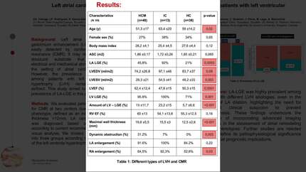
A total of 99 patients were included with final CMR diagnoses of hypertrophic cardiomyopathy (HCM) in 48, infiltrative cardiomyopathy (IC) in 13, and hypertensive cardiomyopathy (HC) in 38. LA-LGE was present in 42 patients (41%). The prevalence of LA cardiomyopathy was significantly different by etiology: 45.8% in HCM, 92.3% in IC, and 21.1% in HC (p:0.0003). Age was significantly different between groups (51.3 vs 63.4 vs 59; p:0.002, respectively) and non-significant differences were found in other clinical features such as female sex, and body mass index. Furthermore, there were significant differences regarding LVEF (62.4% vs 47,8% vs 50.3%; p:<0.001), amount of LV-LGE (13% vs 23.2% vs 5.7%; p:0.001), maximal wall thickness (19.8mm vs 15.5mm vs 12.5mm; p:<0.001), and dynamic obstruction (15% vs 1% vs 0%; p: 0.003, respectively). There were no differences in LA dilation between the three groups (91.6% vs 100% vs 84.2%; p:0.22, respectively). Notably, LA-LGE was detected in 46.1% of patients with LA enlargement but also in 10% of those with normal LA size.
LA-LGE was highly prevalent among patients with different LVH etiologies, even in the absence of LA dilation, highlighting the need for heightened clinical suspicion to prevent underdiagnosis. These findings underscore the importance of incorporating advanced imaging techniques in the assessment of atrial remodeling in LVH phenotypes. Further studies are needed better to define its pathophysiological significance and potential prognostic implications. Characteristics of total population LA-LGE in LVH cardiomyopathies
Contributors


A G Villa
Author
M L Sanchez
Author
M A Bajana
Author
A M Larriva
Author
P A Morejon
Author


A Baranchuk
Author
You may be interested in