Prevalence of inherited cardiomyopathies in patients with sub-epicardial scar on cardiac magnetic resonance imaging
European Heart Journal


Abstract
Pathogenic or likely pathogenic (P/LP) genetic variants, particularly in desmoplakin (DSP), are increasingly recognised causes of arrhythmogenic cardiomyopathy. Diagnosis relies on cardiac magnetic resonance imaging (CMR) and genetic testing. Sub-epicardial late gadolinium enhancement (LGE) is a characteristic CMR finding in DSP cardiomyopathy, but other risk-predictive imaging features are unidentified.
Determine the prevalence of P/LP cardiomyopathy genetic variants in patients with sub-epicardial LGE and delineate imaging predictors that stratify high risk individuals that would most benefit from genetic testing.
Consecutive patients who underwent CMR between 2020 and 2024 at a quaternary centre, with two or more segments of sub-epicardial LGE (without a diagnosis of sarcoid, amyloid or hypertrophic cardiomyopathy), were offered prospective genetic testing with standardised cardiomyopathy panel. CMR volumetric, tissue and contrast characteristics were compared between gene positive and negative groups.
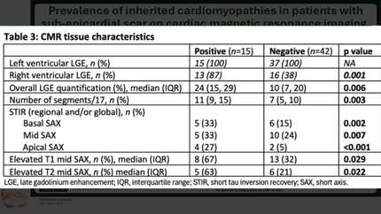
Of 133 patients identified, 52 underwent genetic testing. Nineteen had genetic variants: 15 (29%) P/LP variants (73% desmoplakin, 7% SCN5a, 7% FLNC, 7% TNNT2, 7% TPM1) and 5 (7%) variants of uncertain significance. Baseline characteristics demonstrated in Table. Median left ventricular (LV) ejection fraction was lower in the genotype positive (G+) group (48% (interquartile range (IQR) 41, 55) vs 54% (48, 57), p=0.048), without other volumetric measurements differing between groups. The G+ group was more likely to have evidence of oedema with an elevated short tau inversion recovery (STIR) ratio in the base and apex (5 (50%) vs 6 (17%), p=0.038; 4 (44%) vs 2 (6%), p=0.003). The total LGE burden was significantly higher in the G+ group, with a greater number of affected segments (median: 11 (IQR 9-15) vs 7 (5-10), p=0.002) and degree of hyperenhancement (median: 24% (IQR 15, 29) vs 10% (6, 18), p=<0.001), with more right ventricular involvement (13 (87%) vs 12 (32%), p<0.001). LGE was seen more frequently in most LV segments in the G+ group (p<0.05, Figure). Except there was no difference in the presence of LGE in the basal inferior, basal-mid inferolateral and anterolateral segments and apical lateral segment (segments typically involved in immune mediated myocarditis) (Figure).
Cardiomyopathy genetic variants are prevalent in those with sub-epicardial LGE, with DSP being most common. These results signal a distinct CMR pattern in inherited cardiomyopathies including elevated STIR ratio, increased LGE burden and right ventricular involvement. Interestingly, we demonstrated a patten of global/extensive regional LGE in inherited cardiomyopathy distinct from genotype negative patients that had basal-lateral predominance. These findings may guide clinicians in prioritising patients for genetic testing based on their CMR result. LGE distribution
Contributors

N Sutherland
Author
M Dragas
Author
S Peters
Author
L Salmon
Author
M Cotter
Author
A Harpur
Author
J Martinussen
Author
K Verma
Author
A J Taylor
Author
D M Kaye
Author
H Patel
Author
A Voskoboinik
Author
S J Gutman
Author
You may be interested in