Diagnostic performance of cardiac diffusion tensor imaging in subclinical hypertrophic cardiomyopathy: a comparison with strain analysis
European Heart Journal


Abstract
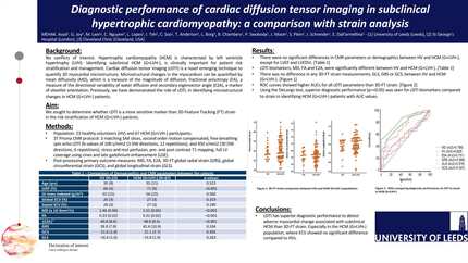
Hypertrophic cardiomyopathy (HCM) is a genetic disease with a prevalence of 1 in 500, characterised by left ventricle hypertrophy. Identifying subclinical HCM, defined as genotype positive phenotype negative (G+LVH-), is clinically important for patient risk stratification and management. We previously demonstrated the role of cardiac diffusion tensor imaging (cDTI) cardiac magnetic resonance (CMR), in identifying microstructural changes in HCM (G+LVH-) patients (1). Myocardial strain with CMR feature-tracking (FT) has prognostic value in overt HCM (2). We sought to determine whether cDTI is a more sensitive marker than 3D-FT strain in the risk stratification of HCM (G+LVH-) patients.
This multi-centre collaboration included 90 subjects: 23 healthy volunteers (HV) and 67 HCM (G+LVH-) patients. All patients underwent 3T CMR imaging with the following protocol: functional imaging, second order motion compensated, free-breathing spin echo cDTI (b-values of 100 s/mm2 (3 DW directions, 12 repetitions), and 450 s/mm2 (30 DW directions, 6 repetitions), pre and post contrast T1 mapping and LGE. Global cDTI analysis was undertaken to derive: mean diffusivity (MD) (a measure of the magnitude of diffusion, high values thought to reflect interstitial fibrosis), fractional anisotropy (FA) (directional variability of water diffusion with low levels thought to be related to collagen infiltration and cardiomyocyte disorganisation), secondary eigenvector angle (E2A) (a marker of sheetlet orientation). cDTI postprocessing was performed using an inhouse Matlab based software with automatic registration. Using Circle Cvi42 software, 3D feature tracking (FT) was derived for global radial strain (GRS), global circumferential strain (GCS), and global longitudinal strain (GLS).
Participants with HCM (G+LVH-) (F:M= 40:27, age 33±11 years) had a LVEF of 71±6%, normal global ECV 27±3%, septal ECV 27±3% and normal LV mass of 54±22 g/m2. Comparing HCM G+LVH- and HV, the cDTI biomarkers were significantly different [MD (x 10-3mm2/s) 1.51±0.05 vs 1.46±0.04 (p< 0.001), FA 0.31±0.02 vs 0.33 ±0.02 (p<0.001) and E2A 48.9°±8.5° vs 40.8°±8.6° (p<0.001)], but strain measurements were not significantly different [GLS 15.9±2.9% vs 16.4±1.6% (p=0.263), GCS 21.1±2.7%, -21.6±1.8% (p=0.505) and GRS 42.4±10.9% vs 39.9±7.9% (p=0.334)]. Global ECV and septal ECV showed no significant differences between G+LVH- and HV subjects (p=0.319 and p=0.190, respectively). ROC curves showed a better diagnostic performance for cDTI biomarkers compared to strain in identifying subclinical HCM with AUC values of: MD 0.769, FA 0.828, E2A 0.731, GLS 0.579, GCS 0.547 and GRS 0.568.
cDTI has better diagnostic performance to detect adverse myocardial change associated with subclinical HCM than 3D-FT strain. cDTI may provide further clinical utility in subclinical HCM.
Contributors

L Lopes
Author
C Soo
Author
T Anderton
Author
L Borg
Author
B Chambers
Author
M E H A K Asad
Author
G Joy
Author
P Swoboda
Author
Leeds Institute of Cardiovascular and Metabolic Medicine Leeds , United Kingdom of Great Britain & Northern Ireland
M Lwin
Author
C Nguyen
Author
J Moon
Author

J Schneider
Author
E Dall'armellina
Author
You may be interested in