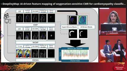
DeepOxyMap: AI-driven feature mapping of oxygenation-sensitive CMR for cardiomyopathy classification
European Heart Journal


Abstract
Cardiovascular disease remains a major global health concern, necessitating advanced, non-invasive diagnostic techniques. [1]Oxygenation-sensitive cardiovascular magnetic resonance (OS-CMR) imaging provides a promising contrast-free approach for myocardial assessment. [2] This study introduces DeepOxyMap, OS-CMR imaging, combined with AI-powered feature mapping, enables myocardial pathology detection without contrast agents. DeepOxyMap classifies and visualize patterns of myocardial scars into ischemic (42), non-ischemic (33), edema (47), and healthy (68) cases, providing the AI-based feature visualization.
Develop DeepOxyMap, a deep learning-based classification model that not only enhances diagnostic precision but also visualizes scar patterns on OS-CMR for the first time, offering a contrast-free alternative to late gadolinium enhancement (LGE).
The dataset comprised short-axis OS-CMR images, LGE, T1, and T2 maps from two independent research cohorts, totaling 160 subjects (43.1 ± 15.3 years, 30.0% female) from the first study and 30 participants (54.93 ± 9.73 years, 50.0% female) from the second study to improve generalizability. A VGG19-based convolutional neural network was employed using transfer learning. Preprocessing included image resizing, normalization, and augmentation with random rotations (±20°, ±40°, ±60°, ±180°) and horizontal flipping (50% probability) to improve model robustness. The dataset was split based on 80% policy and model performance was evaluated using accuracy, precision, recall, and ROC-AUC scores. DeepOxyMap’s feature maps were extracted to visualize pathological patterns and compared against LGE images, serving as the clinical reference.
DeepOxyMap achieved a classification accuracy of 82.0% on the test set, with precision and recall scores of 86.0% and 78.0%, respectively. On the validation set, the model achieved an accuracy of 78.0% and a precision of 84.0%, demonstrating consistency across datasets. Multi-class ROC-AUC analysis showed strong discriminatory power (healthy: 0.94, ischemic: 0.88, non-ischemic: 0.93, edema: 0.98), outperforming ResNet50 (AUC of 0.87) and EfficientNetB0 (0.79) (see Figure 1). Most notably, DeepOxyMap’s feature maps closely aligned with expert-identified LGE, T1, and T2 maps, demonstrating its ability to localize myocardial pathology accurately. For the first time, OS-CMR feature maps exhibited scar patterns comparable to those observed on LGE, reinforcing the potential of this contrast-free imaging approach in clinical settings (see Figure 2).
DeepOxyMap is a clinically viable, AI-driven OS-CMR framework for contrast-free myocardial scar classification and visualization. By generating feature maps that align with LGE, the model enhances diagnostic precision and offers a safer, more accessible alternative to conventional imaging.
Contributors



You may be interested in