Can natural shear wave imaging reveal cardiac dysfunction during follow-up of childhood cancer survivors?
European Heart Journal


Abstract
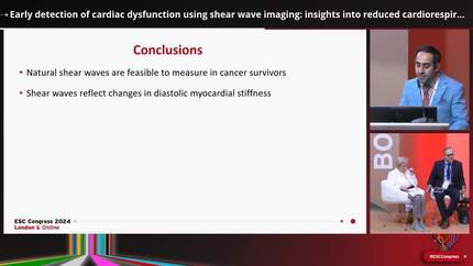
Cardiotoxicity in childhood cancer survivors (CCS) is multifactorial and can further impair the cardiorespiratory fitness (CRF) of survivors, especially during exercise. Cardiopulmonary exercise testing (CPET) with exercise echocardiography can aid in diagnosis, but technical challenges remain. High frame rate (HFR) imaging of natural shear wave (SW) generated by mitral valve closure (MVC) is an innovative parameter for assessing myocardial stiffness, a fundamental element of diastolic function.
To investigate the potential of natural SW velocities measured at rest as a marker of myocardial stiffness to detect reduced cardiorespiratory fitness among CCS during CPET.
A total of 132 subjects; symptom-free 76 CCS (mean age 19.5 ±3.5 years) and 56 matched healthy volunteers (HV) participated in the study. We acquired both conventional as well as high frame rate echocardiographic images (ca. 1300 frames/second) at rest. Participants performed an incremental, symptom-limited cardiopulmonary exercise test on a treadmill to measure peak oxygen consumption (VO2 peak) as a marker of CRF. They were subsequently classified as fit CCS, non-fit CCS and HV. Echocardiographic images were analyzed offline, blinded to the CRF status. SW propagation after MVC was tracked in the parasternal long axis view where an anatomical M-mode was drawn along the ventricular septum and tissue-acceleration maps were extracted. SW after MVC appeared as green bands and their velocities were measured by estimating the slope semi-automatically (figure 1A-C).
In our CCS cohort, the time since treatment ranged from 0.5 to 12.2 years. CCS had significantly lower ejection fraction and global longitudinal strain compared to HV (p<0.01). However, neither parameter correlated with CRF (figure 2A-B). In contrast, SW velocities after MVC measured at rest correlated strongly with VO2 at peak exercise (r=0.67, p< 0.001). Higher SW velocities, indicating increased myocardial stiffness, were associated with reduced CRF (figure 2C). SW velocities were significantly higher among non-fit CCS compared to both fit CCS and HV (4.2 vs 3.5 vs 3.2 m/s respectively, p <0.001) (figure 2D). Additionally among the CCS, a cut-off value of 3.7 m/s for SW velocities measured at rest accurately predict reduced CRF with a 88% sensitivity, 80% specificity, and an accuracy of 83% (AUC= 0.88) (Figure 2E).
Our study data indicate that myocardial stiffness, as determined through SW velocities at rest, is related to cardiorespiratory fitness which explain in part the impaired CRF among childhood cancer survivors. Cardiac natural SW imaging proposes a promising echocardiographic parameter for early detection of subtle cardiac dysfunction.
Contributors


S Verschueren
Author
D Vanrusselt
Author
N Van Ermengem
Author
A Uyttebroeck
Author
L Wouters
Author
N Salerno
Author
M Hornikx
Author
S Pleysier
Author
B Cools
Author
J A N Dhooge
Author
J U Voigt
Author
You may be interested in