Artificial intelligence for automatic echocardiography image view detection and left ventricle segmentation and ejection fraction prediction
European Heart Journal - Cardiovascular Imaging


Abstract
Left-ventricular (LV) border segmentation is key to assess LV size and function. Current assessment has relatively large inter-observer variability, possibly impacting patients care. Artificial intelligence (AI) may permit faster and more reproducible assessment, possibly suitable to be used by non-experts.
To train and test AI algorithms for (i) the identification of views, (ii) the segmentation of the LV in 2, 3 and 4-chamber views, and (iii) the computation of LV ejection fraction (EF).
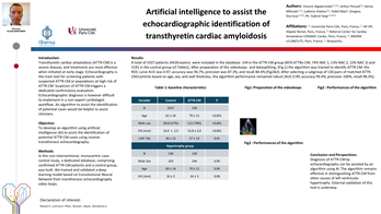
Fifteen thousand echocardiography studies acquired in the Vall d’Hebron University Hospital (VH) for patients care were retrieved and anonymized. LV wall was identified via commercial clinical software in 619 videos with 2-, 3- and 4-chamber views (14082 frames) and 3 masks were created: LV cavity (within endocardial border), overall LV (within epicardial border) and LV wall. Annotated frames were split into independent training (465 videos) and testing (154) sets and performances were evaluated in terms of DICE score. End-diastolic and end-systolic LV volumes of 4-chamber views were identified and used to compute LV EF, which was compared with clinical reports in 488 patients. An external validation on CAMUS dataset was performed.
A total of 6831 videos from 488 VH subjects were available for this analysis (Table 1). AI model identified image views with high accuracy (93%). AI performance on the segmentation of the LV cavity, overall LV and LV wall were satisfactory in 2- (0,86[0,79;0,90], 0,91[0,86;0,93], 0,79[0,74;0,83]), 3- (0,88[0,84;0,91], 0,91[0,90;0,93], 0,81[0,77;0,83]) and 4-chamber (0,90[0,86;0,93], 0,92[0,88;0,94], 0,82[0,79;0,85]) views. Segmentation performances were similar between sexes but were lower in case of atrial fibrillation and in images with low quality. In an external validation in 500 CAMUS patients performances were satisfactory, with DICE score of 0,91[0,87;0,94] and 0,80[0,73;0,84] for whole LV and LV cavity in 2 and 4-chamber views, respectively.
In VH data EF predictions were in line with expected inter-observer reproducibility, with a good linear association (R=0.39, p<0.001) but remarkable underestimation (mean error = 12%). Models performed similarly in the external validation set, with a good linear association (R=0.31, p<0.001) and minimal underestimation (mean error = 2.2%).
AI models can identify echocardiography views, segment the LV in 2, 3 and 4-chamber views and use the segmentation to quantify EF. Care should be taken to avoid biases in AI performances, particularly in case of atrial fibrillation and limited image quality.
Demographic and clinical information
Contributors

A Morales-Galan
Author
J Garrido-Oliver
Author

M Bragulat-Arevalo
Author

A Catala-Santarrufina
Author
G Teixido-Tura
Author
L Galian-Gay
Author
I Ferreira-Gonzalez
Author
J Rodriguez-Palomares
Author

You may be interested in