The crucial role of 18F-fluorodeoxyglucose positron emission tomography/computed tomography in diagnosing pulmonary valve endocarditis in patients after transcatheter pulmonary valve implantation: a case report
European Heart Journal - Case Reports


Abstract
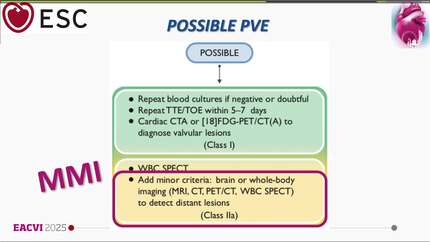
Patients after transcatheter pulmonary valve implantation (TPVI) are at increased risk for infective prosthetic valve endocarditis. Diagnosis of infective endocarditis (IE) following TPVI is particularly difficult due to impaired visualization of the transcatheter pulmonary valve (TPV) with echocardiography [Delgado V, Ajmone Marsan N, de Waha S, Bonaros N, Brida M, Burri H, et al. 2023 ESC guidelines for the management of endocarditis.
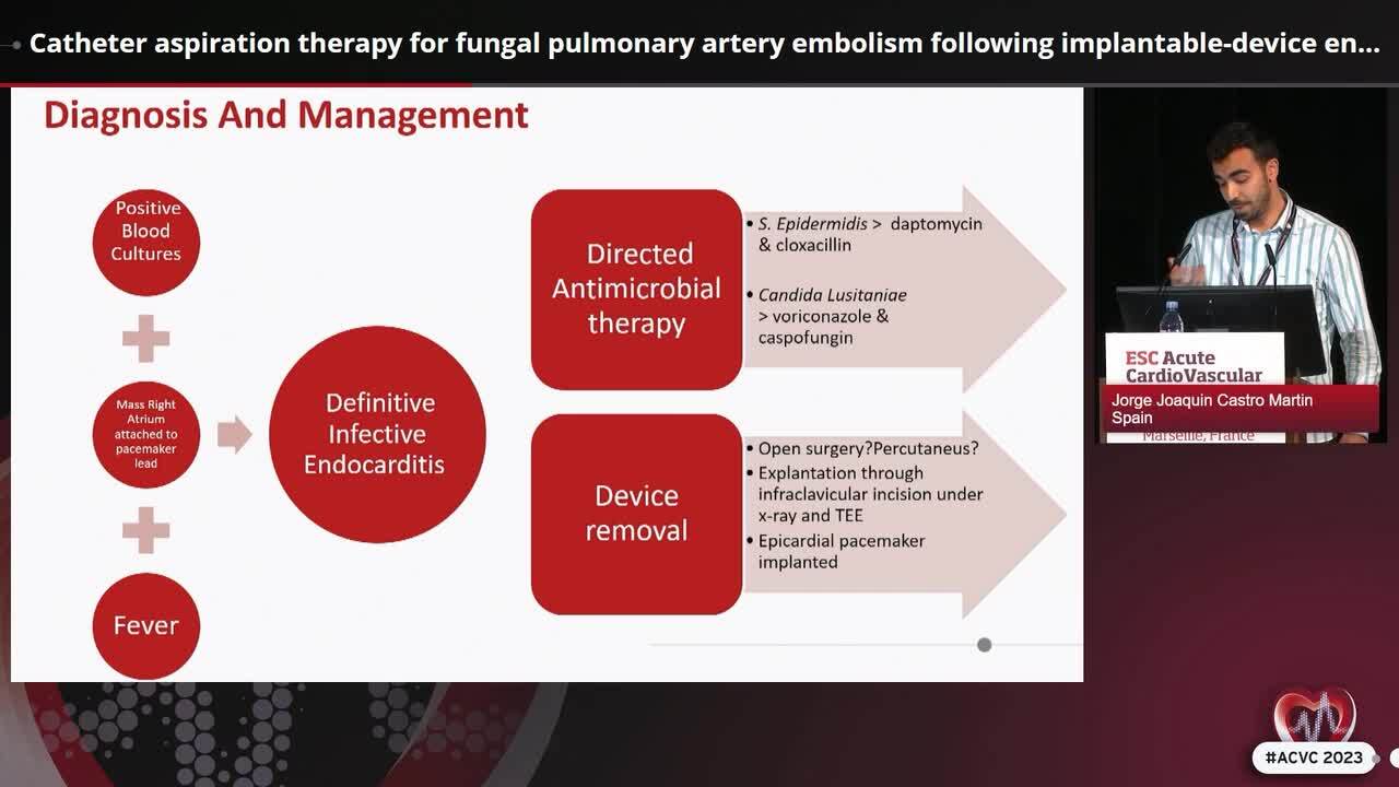
A 22-year-old woman presented to the emergency department with fever and chest pain. Relevant past medical history included a left ventricular outflow membrane resection at infancy, a Ross procedure at the age of 4 with post-operative pacemaker implantation and Melody™ TPVI at the age of 16 because of pulmonary valve stenosis. Blood tests showed elevated inflammatory markers. Transthoracic echocardiography revealed elevated systolic pulmonary artery pressure of 53 mmHg. After 2 days, blood cultures appeared positive for
Transthoracic echocardiography and transoesophageal echocardiography often fail to provide adequate assessment, making 18F-FDG PET/CT crucial for diagnosing TPV IE in this case. Important to notice is the possibility of false-negative and false-positive diagnoses and the radiation exposure, particularly in this young population.
Contributors

Liesbeth Rosseel
Author
Christoph Sinning
Author
Ganesh Gajanan
Author
Marie-Luise Dikou
Author
Mohammed Shahbaaz Khan
Author
Sheetal Vasundara Mathai
Author
You may be interested in