Basal inferior-septal segment is highly susceptible to deformation in the clinical spectrum of transthyretin-derived cardiomyopathy
European Heart Journal


Abstract
The prevalence of transthyretin-derived cardiomyopathy (ATTR-CM) is increasing; however, understanding the morphological and functional manifestations within the left ventricle (LV) across the spectrum of heart failure (HF) remain limited.
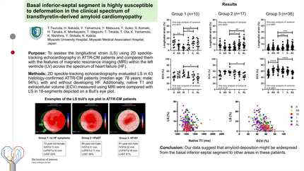
This study aims to assess the longitudinal strain (LS) using 2D speckle-tracking echocardiography in ATTR-CM patients and compared them with the features of magnetic resonance imaging (MRI).
2D speckle-tracking echocardiography evaluated LS in 63 histology-confirmed ATTR-CM patients (median age: 78 years; male: 94%), with and without developing HF. Additionally, native T1 and extracellular volume (ECV) measured using MRI were compared with LS in 18-segments depicted on a Bull’s eye plot.
Patients were grouped based on HF status: Group 1 (no HF symptoms, n=10), Group 2 (HF with preserved LV ejection fraction (LVEF), n=17), and Group 3 (reduced LVEF, n=36). Group 3 exhibited higher levels of brain natriuretic peptide and troponin T, and lower estimated glomerular filtration rate, compared with Group 1 and 2. Echocardiography revealed heightened LV thickness and decreased LVEF in Group 3. LS analysis revealed susceptibility to deformation in the basal inferior-septal segment, persisting even in Group 1. MRI revealed a high average native T1 level with minimal standard deviation (indicative of tissue homogeneity). However, this level increased as distance from the inferior-septal segment grew in the basal and mid-ventricular slices. Contrast images displayed maximal ECV in the basal and mid-ventricular inferior-septum, a phenomenon persisting in Group 1. Segmental LS decline, as assessed by echocardiography, correlated with ECV expansion but not with native T1 values.
2D speckle-tracking echocardiography can identify segmental LS impairment in ATTR-CM patients, potentially reflecting amyloid burden and fibrosis extent. Our data suggest that amyloid deposition might be widespread from the basal inferior-septal segment to other areas.
Contributors


H Nakada
Author
Y Yamamura
Author
Y Matsuura
Author
Y Suiko
Author
S Komaki
Author
H Tanaka
Author
K Moribayashi
Author
T Ideguchi
Author
T Terada
Author
T Ota
Author
K Yamamoto
Author
K Nishihira
Author
Y Shibata
Author
K Kaikita
Author
You may be interested in