Innovative models for cardiotoxicity assessment in human pluripotent stem cell-derived cardiomyocytes that enable clinically relevant treatment regimen
Cardiovascular Research


Abstract
Type of funding sources: Public grant(s) – EU funding. Main funding source(s): This work was supported by the European Research Area Network on Cardiovascular Diseases
807 (2016T092) and Hartstichting (2019T104)
Cardiotoxicity continues to be a significant factor in drug withdrawal, partly due to the unpredictable nature of animal models. Moreover, the limitation of cancer treatment arises from the potential risk of cardiotoxicity. Identifying patients at risk of developing heart symptoms after therapy will strongly contribute to improved treatment possibilities.
Cardiomyocytes derived from human pluripotent stem cells (hPSCs) represent an inexhaustible cell reservoir and hold the potential to provide personalized solutions to address this issue. However, conventional 2D cultures have several limitations, such as tissue complexity and the difficulty to apply repeated dose treatments.
Therefore, we developed an engineered heart tissue (EHT) platform to predict functional aspects of cardiotoxicity. We evaluated the effect of several anti-cancer drugs on cardiomyocyte contractility after repetitive treatment and quantified force of contraction and contraction kinetics.
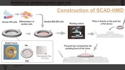
However, these traditional EHTs lack the complexity of diverse cell types. Thus, in a next step, we generated an innovative and user-friendly Heart-on-Chip (HoC) model from hPSC-derived cardiomyocytes, endothelial and smooth muscle cells. Under flow, these miniaturized micro-EHTs (μEHTs) self-organize into a cardiomyocyte core surrounded by an endothelial barrier layer mimicking the cardiomyocyte-endothelial interface as observed in vivo.
Our versatile drug screening systems permit following individual tissues over time. We therefore treated EHTs with DMSO or 1 µM Doxorubicin for 24 hours and monitored tissue contraction force over a time course of 28 days. Doxorubicin treatment initially caused a drop of force, but demonstrated a recovery of force and kinetics 10 days after treatment. To recapitulate a clinically relevant intermittent treatment regime, a second treatment of 1 µM Doxorubicin was administered 21 days after the first dose and showed a similar decrease in contraction force compared with the first exposure to the drug.
μEHTs cultured under flow exhibit improved contractile performance and conduction velocity when compared to static culture conditions. Importantly, the presence of an endothelial barrier delayed the cardiotoxic effect of drugs, indicating that endothelial cells form a barrier mimicking the systemic delivery route of drugs to the heart.
Our systems are characterized by increased physiological accuracy and enable the study of hPSC-cardiomyocyte recovery and the effects of accumulated dose after multiple dosing, allowing individualized cardiotoxicity evaluation. In the future, we can further increase the complexity by including multi-organ-on-chip approaches, as for example heart-liver-on-chip to revolutionize the screening of therapeutic targets and the prediction of drug efficacy.
Contributors

C Cofino-Fabres
Author
R H Slaats
Author
J Rivera Arbelaez
Author
S A Ten Den
Author
M C Ribeiro
Author
You may be interested in