Impact of phase-correction of late-gadolinium enhancement images on quantification of scar metrics and in-silico VT-modelling in patients with an ischemic cardiomyopathy
EP Europace Journal


Abstract
Late gadolinium enhancement (LGE) is used to perform tissue characterization and has become the cornerstone to guide treatment of ventricular tachyarrhythmia (VT). Traditionally, LGE acquisition is reconstructed in two distinct forms: non-phase corrected magnitude (MIR) and phase-corrected inversion recovery (PSIR) images. However, the influence of MIR vs. PSIR on the quantification of scar metrics and the repercussion on estimating myocardial arrhythmogenicity have not been comprehensively explored.
This study investigated the influence of PSIR and MIR LGE on the quantification of scar metrics in a randomly selected cohort of patients with ischemic cardiomyopathy who had undergone imaging prior to ICD implantation and had received device therapy during follow-up. In addition, this study assessed how these image variations affect functional estimation of arrhythmogenicity using computational modelling.
ADAS 3D LV was used to generate scar maps from 2D PSIR and MIR LGE images in 10 patients. The extent of variation in scar-characteristics was evaluated by quantifying the borderzone (BZ), scar core, and conduction corridors. Electrical simulations were performed on the patient-specific anatomical models using the Virtual Induction and Treatment of Arrhythmias (VITA) framework. VITA was used to assess the electrical vulnerability of potential reentrant channels and provided the number of inducible VTs and their corresponding round-trip-time (RTT), an equivalent of VT cycle-length.
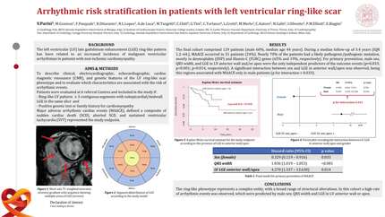
Scar quantification using PSIR LGE resulted in a significantly larger BZ (22.1±23.7g vs. 6.2±2.7g, p <.001) and scar core (43.5±21.5g vs. 10.5±4g, p =.047). Furthermore, both the absolute number (6.8±4 vs. 3.4±1.5, p = .031) and weight (13±13.1g vs. 1.4±0.8g, p =.018) of conduction corridors were also higher using PSIR. VTs were inducible in 8 PSIR-models and 7 MIR-models.
Despite large differences in scar characteristics, VITA metrics were not significantly different between PSIR and MIR derived models. Although, the absolute number of inducible VTs were larger for PSIR (6.2±5.4 vs. 3.4±3.1), non-parametric testing using Wilcoxon signed-rank test did not demonstrate a significant difference (Z = -1.334, p =.139). A comparable, non-significant difference was observed for mean- (117±78 vs. 79±60, p =.257) and max-RTT (181±141 vs. 120±103, p =.294) between PSIR and MIR derived models.
PSIR resulted in significantly larger BZ, scar core area, and conduction corridors. While the absolute, VITA-derived estimation of myocardial arrhythmogenicity, including inducible VTs and RTT, were higher in the PSIR-based quantification, these differences did not reach statistical significance. These results highlight important differences between two clinically applied image reconstruction techniques for the assessment of arrhythmogenic substrate, emphasizing the need for additional investigation and refinement of scar quantification techniques.
Contributors




You may be interested in