Assessing left atrial intramyocardial fat infiltration from computerized tomography angiography in patients with atrial fibrillation
EP Europace Journal


Abstract
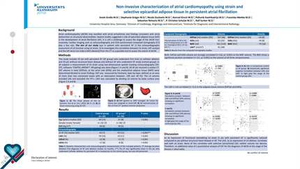
Epicardial adipose tissue might promote atrial fibrillation (AF) in several ways, including infiltrating the underlying atrial myocardium. However, the role of this potential mechanism has been poorly investigated. The aim of this study is to evaluate the presence of left atrial (LA) infiltrated adipose tissue (inFAT) by analysing multi-detector computer tomography (MDCT)-derived three-dimensional (3D) fat infiltration maps and to compare the extent of LA inFAT between patients without AF history, with paroxysmal, and with persistent AF.
Sixty consecutive patients with AF diagnosis (30 persistent and 30 paroxysmal) were enrolled and compared with 20 age-matched control; MDCT-derived images were post-processed to obtain 3D LA inFAT maps for all patients. Volume (mL) and mean signal intensities [(Hounsfield Units (HU)] of inFAT (HU −194; −5), dense inFAT (HU −194; −50), and fat-myocardial admixture (HU −50; −5) were automatically computed by the software. inFAT volume was significantly different across the three groups (
Persistent forms of AF are associated with greater degree of LA intramyocardial adipose infiltration, independently of body mass index. Compared to controls, AF patients present higher LA inFAT volume at left and right superior pulmonary vein antra.
Contributors


Giulio Falasconi
Author
David Soto-Iglesias
Author

Diego Penela
Author
José Alderete
Author
Daniel Viveros
Author
Aldo Francisco Bellido
Author
Paula Franco-Ocaña
Author
Fatima Zaraket
Author
Darío Turturiello
Author


You may be interested in