Role of molecular imaging in the diagnosis of prosthetic aortic valve endocarditis by Bacillus licheniformis: a case report
European Heart Journal - Case Reports


Abstract
Infective endocarditis is a challenging diagnosis that usually requires cardiovascular image confirmation as part of the approach. 18F-Fluorodeoxyglucose positron emission tomography/computed tomography (18F-FDG-PET/CT) is an imaging technique more sensible for the diagnosis of prosthetic valve endocarditis (PVE) when echocardiography is inconclusive.
We present the case of a 35-year-old man who had a previous Bentall–De Bono procedure 4 years prior that included biological, national institute of cardiology (INC)-type, locally manufactured aortic valve replacement and woven Dacron tube graft implantation in the ascending aorta. He was admitted because of dyspnoea, oedema, fever, and syncope. A complete auriculoventricular blockade was diagnosed, requiring cardiac pacing. Also, infective endocarditis (IE) was suspected. Blood cultures showed the isolation of
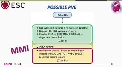
Although the other imaging modalities were negative, the high clinical suspicion made it mandatory to continue the study protocol, remarking on the utility of 18F-FDG-PET/CT on patients categorized as having ‘possible’ endocarditis, as in our patient.
Contributors

Juan Carlos Plata-Corona
Author
Araceli González-Ortiz
Author
Mauricio García-Cárdenas
Author
Nilda Espinola-Zavaleta
Author
Erick Alexanderson-Rosas
Author
Isabel Carvajal-Juárez
Author
Luca Arcari
Author
Michael ZL Zhu
Author
Marianna Garcia-Saldivia
Author
Emmanouil Mantzouranis
Author
Gonçalo Costa
Author
You may be interested in