The role of four-dimensional computed tomography in transcatheter aortic valve replacement prosthesis endocarditis with concurrent leaflet thrombosis: a case report
European Heart Journal - Case Reports


Abstract
Transcatheter aortic valve replacement (TAVR) is becoming increasingly utilized for the treatment of severe aortic valvular heart disease. Infective endocarditis of TAVR is rare but associated with higher mortality and morbidity. The potential for leaflet thrombosis following TAVR is also becoming increasingly recognized. Diagnosis of these conditions on echocardiography can be challenging due to prosthesis artefact.
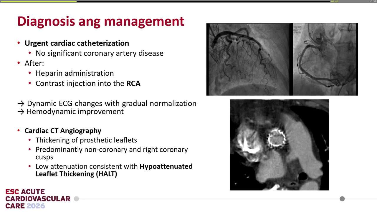
An 84-year-old man with a previous transcatheter aortic valve replacement presented with a febrile illness and bacteraemia. Transthoracic and transoesophageal echocardiography demonstrated high transvalvular gradients with features of prosthesis endocarditis, though leaflet morphology could not be fully assessed due to prosthesis artefact. Four-dimensional computed tomography revealed hypo-attenuated leaflet thickening with reduced leaflet motion, consistent with prosthesis leaflet thrombosis. The patient was successfully treated with antibiotics and anticoagulation, with resolution of the infection and normalization of the transvalvular gradient after 6 weeks.
Echocardiography should be the first-line investigation for assessing leaflet morphology in suspected prosthetic valve endocarditis or leaflet thrombosis but its accuracy may be limited by artefact. Our case highlights that four-dimensional computed tomography provides further evaluation of prosthesis leaflet morphology/motion, providing valuable diagnostic information.
Contributors

Nancy Khav
Author
Hashrul N Rashid
Author
Adam J Brown
Author
Royal Papworth Hospital NHS Foundation Trust Cambridge , United Kingdom of Great Britain & Northern Ireland
Harun Kundi
Author
Carlos Minguito Carazo
Author
Richard Alexander Brown
Author
Kiran Sarathy
Author
You may be interested in