Left atrial intramural haematoma: a fatal complication of cardiac amyloidosis—a case report
European Heart Journal - Case Reports


Abstract
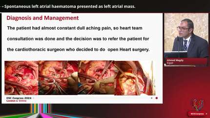
Spontaneous left atrial intramural hematoma (LAIH) is extremely rare and there are only two cases of spontaneous LAIH involving cardiac amyloidosis (CA) reported in literature. In both cases, LAIH rapidly compromised hemodynamic stability proving to be a rare yet fatal complication.
An 83-year-old man presented with cardiogenic shock. Electrocardiogram showed complete atrioventricular block, and echocardiogram revealed severe hypokinesis and left ventricular hypertrophy. Coronary angiography revealed no significant coronary stenosis and tissue biopsy was taken from the left ventricle. The patient was intubated, placed on extracorporeal membrane oxygenation with intra-aortic balloon pump and temporary pacemaker, and admitted to ICU. Day 6 of admission, he became hemodynamically unstable, and presented with atrial fibrillation. Transesophageal echocardiography showed a newly formed large mass in the left atrium. Day 11 of admission, the patient passed away. Autopsy revealed cardiac amyloidosis and showed the mass to be a left atrial intramural hematoma. Diffuse amyloid deposits were found in the myocardium as well as the blood vessel walls of the region surrounding the LAIH.
LAIH is a rare yet fatal complication of CA. Autopsy revealed diffuse amyloid deposits within the left atrium may lead to left atrial fragility and contribute to development of LAIH. LAIH should be considered as an important differential diagnosis in the setting of a rapidly growing left atrial mass, and in hemodynamic instability in patients with CA.
Contributors

Rika Terashima
Author
Takashi Nakagawa
Author
Hisao Hara
Author
Yukio Hiroi
Author
Davide Stolfo
Author
Andreas Giannopoulos
Author
Ana Hurtado-Doce
Author
Annachiara Pingitore
Author
Nicholas Weight
Author
Abdullah Abdullah
Author
You may be interested in