Point-of-care echocardiography screening for hypertrophic cardiomyopathy using automated deep-learning analysis
European Heart Journal - Digital Health


Abstract
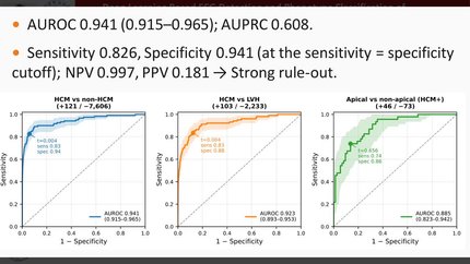
Hypertrophic cardiomyopathy (HCM) remains underdiagnosed due to limited access to expert imaging. We developed and validated a deep-learning (DL)-based echocardiographic model adaptable to point-of-care ultrasound (POCUS) for scalable HCM screening.
We retrospectively analysed 134 956 expert transthoracic echocardiograms (TTE) from 73 598 patients at Sheba Medical Center (2007–2022). A TTE-trained DL model integrating structural features and temporal motion patterns from parasternal long-axis and apical four-chamber views estimated HCM probability. Performance was evaluated in an independent test cohort and clinical subgroups. External validation used bedside POCUS studies from non-cardiologists with handheld devices. The test cohort included 12 096 patients with 119 confirmed HCM cases (prevalence 0.98%; median age 75 years, 57% male). HCM-positive patients showed increased expert TTE-measured septal (1.67 [1.5, 2.0] vs. 1.01 [0.9, 1.19] cm) and posterior wall thickness (1.1 [1.0, 1.3] vs. 0.9 [0.8, 1.0] cm) (
This DL-based model identifies HCM and demonstrates feasibility for POCUS screening, supporting earlier detection and broader diagnostic access.
Contributors

Nour Karra
Author
Yarin Klempfner
Author
Viana Copeland
Author
Michael Fiman
Author
Harel Doitch
Author
Roei Merin
Author
Robert Klempfner
Author
Ehud Schwammenthal
Author
Michael Arad
Author
Elad Maor
Author
You may be interested in