Voluminous calcified pericardial cyst with severe right ventricular compression: a multimodality imaging case report
European Heart Journal - Case Reports


Abstract
Pericardial cysts are rare lesions that are typically asymptomatic. However, when large or calcified, they may compromise cardiac function.
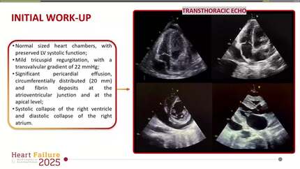
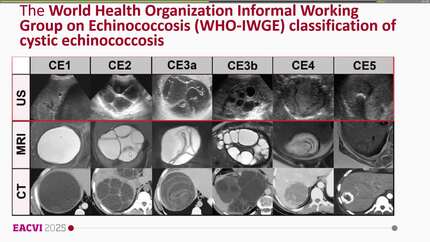
We report the case of a 57-year-old man with progressive exertional dyspnoea. Multimodality imaging revealed a large calcified pericardial cyst causing significant right ventricular compression and interventricular dependence. Given his history of prolonged residence in Central America, a parasitic aetiology was suspected. Surgical drainage with partial resection was performed under cardiopulmonary bypass. Histology confirmed a parasitic process. Postoperative recovery was uneventful, and the patient was discharged on antiparasitic therapy with full clinical resolution.
This case underscores the crucial role of multimodality imaging in the assessment of unusual pericardial disease and highlights the importance of considering parasitic aetiologies in patients with relevant epidemiological risk factors.
Contributors

Alessandra Volpe
Author
Anna Apostolo
Author
Maria Elisabetta Mancini
Author
Linda Bussini
Author
Gianluca Pontone
Author
Sarandeep Kaur Marwaha
Author
Alessandro Palmieri
Author
Sumit Sohal
Author
Deepti Ranganathan
Author
You may be interested in