Deep learning-derived biological age from preoperative chest radiographs predicts mortality following surgical and transcatheter procedures for structural heart disease beyond EuroSCORE II
European Heart Journal - Digital Health


Abstract
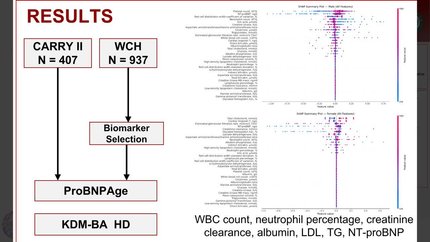
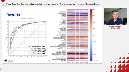
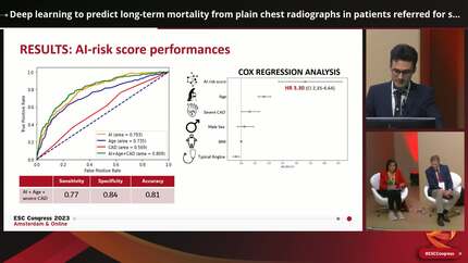
Accurate risk stratification before structural heart disease interventions is essential for clinical decision-making. Traditional risk models, such as the European System for Cardiac Operative Risk Evaluation II (EuroSCORE II) and Society of Thoracic Surgeons Predicted Risk of Mortality (STS-PROM), were designed for surgical patients and show inconsistent performance in transcatheter cohorts. Biological age, reflecting cumulative physiological decline, may offer prognostic value beyond chronological age and established risk scores.
In this retrospective study of 1269 patients [non-transcatheter aortic valve implantation (TAVI)
Biological age distinguished risk across both populations, suggesting that deep learning-based biological age estimation from routine chest radiographs could serve as an automated, accessible complement to existing risk models.
Contributors

Era Stambollxhiu
Author
Miriam Kumpf
Author
Maximilian-Niklas Bonk
Author
Lisa C Adams
Author
Marcus Makowski
Author
Martin Hadamitzky
Author
Markus Krane
Author
Keno K Bressem
Author
Oliver Deutsch
Author
You may be interested in