Validation of a deep learning-based workflow for the interpretation of the echocardiogram in a cardiooncology population
European Heart Journal Supplements


Abstract
Echocardiography is the cornerstone for risk stratification, diagnosis, and monitoring of cancer therapy–related cardiac dysfunction (CTRCD)(1). Artificial intelligence (AI)–guided echocardiography has shown high accuracy and reliability in diverse cardiac populations and may reduce variability while improving workflow efficiency(2, 3). However, this technology has not yet been validated in a dedicated cohort of patients with cancer.
To evaluate the accuracy and reliability of AI-guided echocardiography in assessing left ventricular ejection fraction (LVEF) and additional parameters, compared with conventional echocardiography, in a cardio-oncology population.
This study included patients identified retrospectively from a cardio-oncology registry. Studies, that had already been analysed manually by expert sonographers and reported using AGFA PACS system, were uploaded to the US2.ai platform for automated analysis. The primary outcome was the level of agreement (LoA) between AI-guided and standard echocardiography for LVEF. Secondary outcomes included LoA for additional echocardiographic parameters and LoA between AI- LVEF and 3D LVEF. The performance of the deep learning (DL) algorithm in identifying LVEF <50% was evaluated using the area under the receiver operating characteristic curve (ROC-AUC). Subgroup analyses were performed in predefined populations clinically relevant in cardio-oncology.
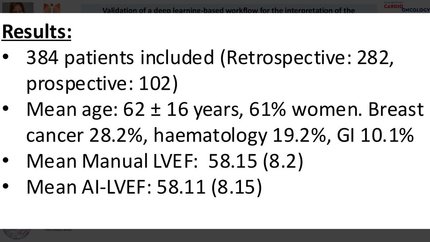
A total of 282 patients were included. Mean age was 60 ± 16 years, and 61% were women. Breast cancer was the most frequent malignancy (30.5%), followed by haematological malignancies (16.7%) and gastrointestinal tumours (10.6%). Manual median 2D LVEF was 60% (IQR: 55-64) and AI-derived LVEF was 59.2% (IQR: 53-64) showing good agreement and correlation (bias: −0.138, SD: 5.38, 95% LoA: −10.7 to 10.4, ICC: 0.791, 95% CI: 0.742–0.831, Spearman ρ: 0.718,), Table 1. Comparison between 3D echocardiography LVEF and AI-derived 2D LVEF showed similar agreement with narrower limits (bias: −0.13, 95% LoA: −9.51 to 9.26). The DL algorithm accurately identified LVEF <50% (ROC-AUC: 0.918, 95% CI: 0.875–0.961), Figure 1. Subgroup analyses demonstrated consistent agreement in patients with breast cancer, body mass index >30, prior radiotherapy and pericardial effusion.
In a large real-world cardio-oncology cohort, AI-guided echocardiography demonstrated strong agreement with conventional echocardiography for LVEF assessment and high accuracy for detecting clinically relevant LV dysfunction. Performance was consistent across key subgroups, supporting the feasibility, reliability, and potential clinical value of integrating DL-based analysis into routine cardio-oncology echocardiographic workflows. Agreement between manual and AI-echo AUC-ROC curve for LVEF<50%
Contributors


V Maharajan
Author
M C Llamedo
Author
J Cervantes
Author
S Ohri
Author
M S Nazir
Author
R Khattar
Author
A R Lyon
Author
You may be interested in