A case report of giant left ventricular outflow tract pseudoaneurysm in a pregnant woman with Loeys–Dietz syndrome: a management conundrum
European Heart Journal - Case Reports


Abstract
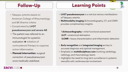
Loeys–Dietz syndrome (LDS) is a rare connective tissue disorder associated with aortic aneurysms and increased surgical complications. Recognizing left ventricular outflow tract (LVOT) pseudoaneurysm after aortic root surgery is challenging but is essential given rupture risk.
A 20-year-old woman with genetically confirmed LDS (TGFBR2-related) and history of elective valve-sparing aortic root replacement for a 4.3 cm aortic aneurysm presented with progressive substernal chest pain and dyspnoea. Transthoracic echocardiography revealed a giant LVOT pseudoaneurysm and mildly reduced left ventricular systolic function. Chest CT angiography confirmed the diagnosis and defined its extent. She underwent urgent surgical repair using a bovine pericardial patch with intraoperative finding of suture-line dehiscence at the aorto-mitral intervalvular fibrosa. Four weeks after redo cardiac surgery, she was found to be 7 weeks pregnant and, after counselling regarding her extremely high-risk status, opted to continue the pregnancy.
This case illustrates the complexities of surgical intervention in LDS, where tissue fragility and comorbidities increase perioperative risk and complicate management. Close postoperative TTE may enable early detection of LVOT pseudoaneurysm after aortic surgery. A low threshold for chest CTA is key for distinguishing true from pseudoaneurysm. Shorter surveillance intervals may be warranted in high-risk patients with risk factors such as tissue fragility, complicated postoperative course, or chest wall deformity.
Contributors

James Rice
Author
Esosa Odigie-Okon
Author
Anastasia Vamvakidou
Author
Konstantinos Bitos
Author
Andrea Papa
Author
Deepti Ranganathan
Author
You may be interested in