Coronary–bronchial artery fistula arising from the sinoatrial nodal artery in a patient with bronchiectasis: a multimodality imaging case report
European Heart Journal - Case Reports


Abstract
Coronary–bronchial artery fistulas are rare vascular anomalies between the coronary and bronchial circulations. They are most often detected incidentally and may be congenital or acquired, particularly in the setting of chronic pulmonary disease. Origin from the sinoatrial nodal artery is exceptional. If haemodynamically significant, these communications may lead to a coronary steal phenomenon and represent a potential cause of myocardial ischaemia. Recognition is also crucial prior to bronchial artery embolization to avoid inadvertent coronary complications. Multimodality imaging plays a key role in their diagnosis and characterization.
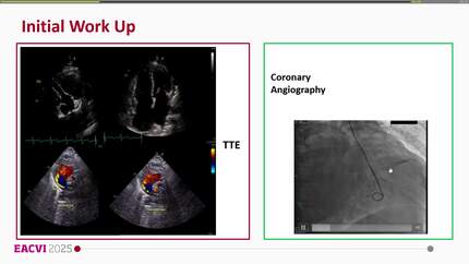
A 58-year-old man with psoriasis presented with atypical chest pain. Exercise stress echocardiography was electrically equivocal, prompting invasive coronary angiography. No significant coronary artery disease was identified. However, contrast injection into the proximal right coronary artery demonstrated a prominent atrial branch with opacification of the right pulmonary parenchyma. Coronary computed tomography angiography revealed a retroaortic sinoatrial nodal artery originating from the proximal right coronary artery. Its left branch communicated with a hypertrophied bronchial artery, which followed a tortuous trajectory towards the right middle lobe. Chest computed tomography demonstrated cylindrical and varicose bronchiectasis in the right middle and lower lobes. No haemoptysis occurred.
This case illustrates a rare coronary–bronchial artery fistula arising from the sinoatrial nodal artery in association with bronchiectasis. Multimodality imaging enabled accurate anatomical characterization and suggested possible haemodynamic relevance. Such fistulas may represent a potential cause of myocardial ischaemia and should be recognized both in the evaluation of chest pain and prior to interventional procedures.
Contributors

Maria Nieves Montoro-Lopez
Author
Gisela Feltes
Author
Ángel Domínguez Álvarez
Author
Iván J Núñez-Gil
Author
Raheel Ahmed
Author
Ugur Canpolat
Author
Mattia Alberti
Author
Deepti Ranganathan
Author
You may be interested in