Trans-esophageal and trans-arteriel pulsed-field delivery to target epicardium in a hybrid in-vitro model
EP Europace Journal


Abstract
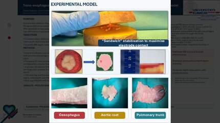
Endocardial ablation may occasionally fail to achieve sufficient lesion depth for epicardial atrial substrates. Pulsed-field ablation (PFA) could enable epicardial targeting from extracardiac structures, but the impact of barrier tissues is unclear.
To quantify how esophagus, aortic root, and pulmonary trunk modulate PFA lesion formation compared with direct delivery.
In-vitro potato slices were overlaid with animal tissues. Energy was delivered using a multielectrode bipolar PFA catheter across three application subsets. We measured depth of irreversible electroporation (IRE), total lesion depth, and IRE surface area. Groups: control and three barriers; 60 lesions in total (n=15 per group; n=5 per subset).
Barrier tissues reduced IRE depth versus control (4.05±1.27 mm): esophagus 2.04±0.33 mm, aorta 1.84±0.74 mm, pulmonary trunk 2.61±0.67 mm (all p<0.001). Total depth was comparable for esophagus (6.65±1.47 mm, p=1.0) and pulmonary trunk (5.55±0.96 mm, p=0.263) but lower for aorta (4.32±1.15 mm, p=0.001). Relative to control IRE area (1,560.48±180.31 mm²), aorta was smaller (1,226.82±247.43 mm², p<0.001), pulmonary trunk similar (1,537.69±125.95 mm², p=0.991), and esophagus larger (1,782.81±264.44 mm², p=0.028).
PFA delivered from extracardiac structures can reach epicardial targets in an ex-vivo model, with tissue-specific attenuation. Oesophageal tissue broadened the affected area with shallower IRE depth, whereas the aortic wall showed greater attenuation of both depth and area.
Contributors

L Dippel
Author
J Minners
Author
J Steinfurt
Author
J Mueller
Author
D Westermann
Author
H Lehrmann
Author
T Arentz
Author
M Eichenlaub
Author
You may be interested in