Three-month 7-Tesla brain MRI after atrial fibrillation ablation detects no new ischaemic lesions: a prospective pilot study
EP Europace Journal


Abstract
Catheter ablation for atrial fibrillation (AF) can generate silent cerebral lesions (SCLs). Standard 3- Tesla MRI may miss very small lesions; whether ultra-high-field 7-Tesla (7T) improves detection at follow-up is unknown.
To evaluate 7T brain MRI for the detection of new ischaemic or haemorrhagic lesions three months after AF ablation and to assess changes in cognition.
We prospectively enrolled patients with persistent or paroxysmal AF without MRI contraindications. All underwent 7T brain MRI and Montreal Cognitive Assessment (MoCA; 0–30) before ablation and at 3 months. Rhythm follow-up included 24-hour Holter at 3 months. Ablation energy was cryoballoon, radiofrequency, or pulsed-field ablation with standard lesion sets. Primary outcome: new ischaemic lesions on 7T at 3 months. Secondary outcomes: haemorrhagic lesions, change in MoCA, and AF recurrence.
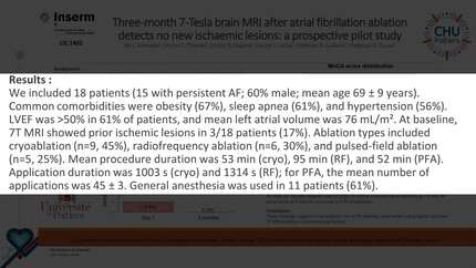
We included 21 patients (18 persistent; 67% male; mean age 66.3 ± 10.2 years). Common comorbidities were obesity (81%), hypertension (66.7%), and sedentary lifestyle (71.4%). LVEF was >50% in 71.4%; mean left atrial volume 170.9 mL (79.1 mL/m²). At baseline, 7T MRI showed prior ischaemic lesions in 2/21 (9.5%).
Ablation energy was cryoablation in 38.1% (n=8), radiofrequency in 23.8% (n=5), and pulsed-field ablation in 23.8% (n=5). Procedure duration was 60 ± 17.3 minutes; total ablation time averaged 1,019 s (cryo), 1,404 s (RF), and 110 s (PFA).
Primary outcome: no new ischaemic lesions were detected on 7T MRI at 3 months (0/21; 0%), irrespective of ablation energy. Secondary outcomes: MoCA was 24.5 ± 2.2 pre-ablation and 24.4 ± 3.6 at 3 months; AF recurrence at 3 months occurred in 27%.
In this prospective pilot, 7T brain MRI at 3 months post-AF ablation detected no new ischaemic lesions across cryo, RF, and PFA energy sources, and cognitive performance was unchanged. These findings suggest that persistent cerebral injury detectable at 3 months is uncommon. Ongoing work will assess early (≤24 h) 7T MRI to characterise acute lesions and explore cerebral perfusion.
Contributors

You may be interested in