Cardiac tomography predictors of functional left atrium substrate in candidates to atrial fibrillation ablation
EP Europace Journal


Abstract
In recent years, interest has grown in the functional substrate underlying atrial fibrillation (AF), particularly slow conduction velocity (SC) [1-3].
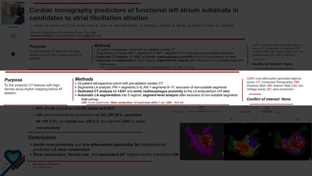
To investigate the relationship between low-attenuation epicardial adipose tissue (I-EAT), aortic root and esophagus proximity to the left atrium (LA), and conduction velocities and voltage on high-density electroanatomic maps.
We retrospectively enrolled 22 consecutive patients undergoing AF ablation who had both cardiac CT and high-density LA mapping in sinus rhythm. Images were processed with dedicated software to identify I-EAT and aortic root and esophagus proximity to the LA endocardium (<1 mm). Predefined HU thresholds from prior literature were applied. [4] The LA was divided into segments, defined automatically according to a previously described method to ensure comparability: roof, posterior wall, septum, anterior wall and lateral wall. [3] Non suitable segments were excluded and a segment-level analysis was performed. Sinus-rhythm activation maps were acquired using a window from P-wave onset to QRS onset, with 12-ms isochrones. Slow conduction was defined as ≥3 isochrones within a 1-cm radius (propagation velocity <27 cm/s). Low voltage (LVA) was defined as bipolar potential <0.5 mV. [2, 5].
258 segments from 22 patients were analyzed. 176 segments (68%) showed SC. Most SC zone were found in anterior wall (65/176; 36.9%). On univariate analysis, SC was associated with persistent AF (p=0.021), low-attenuation epicardial adipose tissue (I-EAT; p<0.001), and aortic-root proximity <1 mm (p=0.034), but not esophageal proximity <1 mm (p=0.718). In multivariable logistic regression, I-EAT (OR 3.69, 95% CI 1.9–7.0; p<0.001), aortic-root proximity (OR 3.3, 95% CI 1.3–8.4; p=0.01), and persistent AF (OR 2.1, 95% CI 1.1–3.9; p=0.02) remained independently associated with SC. [Figure 1] Eighty-three segments (32%) demonstrated low-voltage areas (LVA); 82/83 (99%) colocalized within SC zones (p<0.001). At univariate analysis, LVA was associated with I-EAT (p=0.020), aortic-root proximity (p=0.019), persistent AF (p<0.001), and female sex (p=0.03). In multivariable analysis, LVA was independently associated with SC (OR 59.3, 95% CI 7.9–447.4; p<0.001), persistent AF (OR 4.31, 95% CI 2.2–8.6; p<0.001), and female sex (OR 2.3, 95% CI 1.1–4.7; p=0.03), but not I-EAT (OR 1.62, 95% CI 0.8–3.4; p=0.20) and aortic-root proximity (OR 1.6, 95% CI 0.7–3.9; p=0.38). [Figure 1]
Aortic root proximity and low-attenuation epicardial fat are independently associated with SC in the LA. Slow conduction, female sex, and persistent AF are independently associated with LVA.
Contributors


M Parollo
Author
A N D R E A Di Cori
Author
R De Lucia
Author
A Canu
Author
M Giannotti Santoro
Author
V Barletta
Author
L Segreti
Author
S Sbragi
Author
G Grifoni
Author
S Viani
Author
G Zucchelli
Author
You may be interested in