Intravascular lithotripsy for recurrent restenosis caused by a calcified nodule in a saphenous vein graft: a case report
European Heart Journal - Case Reports


Abstract
Saphenous vein graft (SVG) lesions occurring in the chronic phase after coronary artery bypass graft (CABG) surgery are typically characterized by friable atherosclerotic plaques, whereas stenosis due to calcified nodules (CNs) is rare. Intravascular lithotripsy (IVL) is an effective treatment for severely calcified coronary artery lesions, but its application in SVG lesions remains limited.
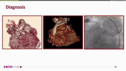
An 84-year-old man with a history of two prior CABG procedures was referred to our hospital with angina pectoris. Coronary angiography revealed severe stenosis in the proximal segment of the SVG to the left anterior descending artery. Intravascular ultrasound demonstrated that a large CN was responsible for severe stenosis at the lesion site. Initial treatment consisted of drug-coated balloon (DCB) therapy following predilatation with a cutting balloon. Because restenosis occurred 9 months later, repeat DCB therapy following more aggressive lesion preparation with high-pressure non-compliant balloon dilatation was performed. Despite repeated DCB treatments, angiographic restenosis recurred 17 months after the initial intervention. Optical coherence tomography (OCT) confirmed a persistent CN, and IVL was performed using a 3.0 × 12 mm ‘Shockwave’ IVL balloon to modify the CN, followed by scoring balloon dilatation and implantation of a 3.5 × 28 mm drug-eluting stent. Final OCT demonstrated improved lumen expansion compared with previous interventions, with a favourable procedural outcome.
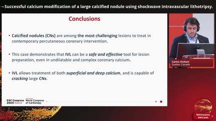
This case highlights the rarity of CNs in SVG lesions and suggests that IVL may be a reasonable treatment option for CNs in this setting.
Contributors

Daisuke Irie
Author
Hibiki Iwakoshi
Author
Atsushi Hosokawa
Author
Yoshio Sasaki
Author
Ryoji Kitamura
Author
Dzan Horozic
Author
Antonios Karanasos
Author
A Shaheer Ahmed
Author
Deepti Ranganathan
Author
You may be interested in