Primary synovial sarcoma of the pericardium diagnosed after multiple biopsies following recurrent episodes of haemorrhagic pericardial effusion following infection: a case report
European Heart Journal - Case Reports


Abstract
Primary malignant pericardial tumours are extremely rare, and synovial sarcoma arising from the pericardium is an exceptionally uncommon entity with poor survival. Diagnosis is often delayed because initial pericardial biopsies can be non-diagnostic, and distinguishing between inflammatory disease and malignancy may be challenging.
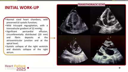
An 11-year-old boy developed recurrent fever and chest pain 15 days after Coronavirus disease 2019 infection. Echocardiography demonstrated pericardial effusion, and pericarditis was suspected. Despite drainage of haemorrhagic effusion, pericardial biopsy showed only fibrous thickening. Two months later, recurrent effusion and haematoma enlargement prompted repeat evaluation, but multimodal imaging including computed tomography, magnetic resonance imaging, and catheterization failed to identify the aetiology. At 3 months, worsening haematoma around the left ventricular posterior wall necessitated surgical excision. The mass was tightly adherent to the pericardium, precluding complete resection. Histology revealed spindle-shaped tumour cells, and molecular testing confirmed SS18::SSX fusion, leading to a diagnosis of synovial sarcoma. 18F-fluorodeoxyglucose positron emission tomography/computed tomography (18F-FDG PET/CT) showed pericardial uptake without distant disease. The patient underwent chemotherapy, radiotherapy, and high-dose chemotherapy with autologous stem cell rescue. He remained in remission for 18 months, but subsequently developed brain metastases and died.
This case highlights the diagnostic difficulty of primary pericardial synovial sarcoma. Initial biopsy may be falsely negative, and repeated sampling directly from the lesion is crucial when a cardiac tumour is suspected. Furthermore, 18F-FDG PET/CT proved valuable not only for diagnostic assessment but also for therapeutic monitoring. In summary, repeated targeted biopsy and multimodality imaging are essential for accurate diagnosis and management of this rare cardiac malignancy.
Contributors

Masayoshi Mori
Author
Yoichiro Ishii
Author
Yosuke Okada
Author
Makoto Takeuchi
Author
Hisaaki Aoki
Author
Takeshi Kitai
Author
Ahmet Taha Sahin
Author
Nidhish Tiwari
Author
Deepti Ranganathan
Author
You may be interested in