Acute coronary syndrome due to papillary fibroelastoma detected by intravascular ultrasound imaging: a case report
European Heart Journal - Case Reports


Abstract
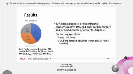
Papillary fibroelastomas (PFEs) are rare benign cardiac tumours that most commonly affect the left-sided heart valves, especially the aortic valve. Although frequently asymptomatic, PFEs can cause embolic events, arrhythmias, or coronary artery occlusion.
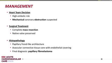
A 57-year-old woman with no significant medical history presented with acute chest pain and was urgently transferred to our hospital. Electrocardiography on admission showed ST-segment elevation in aVR with diffuse ST-segment depression, consistent with acute coronary syndrome (ACS). While in the emergency department, she developed a refractory ventricular fibrillation storm requiring cardiopulmonary resuscitation, intravenous amiodarone, and repeated defibrillation. Sinus rhythm was not restored, so veno-arterial extracorporeal membrane oxygenation (VA-ECMO) was initiated in the catheterization laboratory, followed by immediate coronary angiography (CAG). CAG revealed an intracoronary mass in the left main trunk (LMT). Intravascular ultrasound (IVUS) identified a heterogeneous, mulberry-like mass occluding the LMT and protruding into the aorta. Given suspicion of a soft tissue tumour near the coronary artery origin, surgical resection was performed. Histopathology confirmed the diagnosis of PFE.
This case highlights the crucial role of IVUS in diagnosing rare causes of ACS, such as PFE-related coronary occlusion. IVUS can differentiate tumour from thrombus, enabling timely surgical intervention and improving clinical outcomes.
Contributors

Hiroto Aikawa
Author
Toru Kameda
Author
Toki Toi
Author
Takanori Ikeda
Author
Milenko Zoran Cankovic
Author
Hibatallah Altaher
Author
Deepti Ranganathan
Author
You may be interested in