A novel mapping strategy for ventricular abnormal activities detection in scar-related ventricular tachycardia
European Heart Journal


Abstract
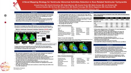
[Background] Estimating the critical isthmus and successfully eliminating delayed potential (DP) and local abnormal ventricular activities (LAVA) form an important strategy for the treatment of scar-related ventricular tachycardia (VT). However, the identification of slow conduction becomes intricate when multiple VTs occur. In our recent work, we have reported a case of VT ablation using the ‘Early Meets Late’ (EML) function in the CARTO system, which highlights differences in local activation time within the total activation time (TAT). 1) Despite the promising approach, the effectiveness of this technique has not yet been fully evaluated.
[Purpose]We aimed to investigate the utility of EML function as a functional substrate map, a technique we have termed the Ventricular Abnormal Activities Detection (VAAD) map.
[Methods] We conducted a retrospective review of 54 cases who underwent scar-related VT ablation using CARTO at our institution between 2019 and 2023. Substrate mapping was performed during sinus rhythm, and in cases of atrioventricular block, mapping was conducted with right ventricular pacing. DP and LAVA were manually marked during the substrate mapping procedure. The critical isthmus of VT was defined as the site where pace-mapping matched all 12 leads or the site of VT termination by ablation. We adjusted the lower threshold of EML ranging from 10% to 30% in 5% increments and determined the consistency between the highlighted site and DP, LAVA, and critical isthmus. (Figure 1) DP, LAVA, and critical isthmuses were counted as a match if they were within 4 mm of the EML highlight site.
[Results] The study population (mean age: 67± 11 years) was 91% male, with an average LVEF of 35 ± 10% and ischemic cardiomyopathy in 56% of cases. The average number of induced VTs was 2. Ventricular mapping was performed in 4 cases of the right ventricle, 54 in the left ventricular endocardial, and 13 epicardial cases (total: 71 maps). Non-inducibility of VT was achieved in 47 cases. Substrate mapping was performed during sinus rhythm in 42 cases and during right ventricular pacing in 12 cases. The average TAT was 201 ms. Of the points obtained (mean: 7,753 points), 3.7% were highlighted at EML30, 7.8% at EML20, and 15.2% at EML10. DP was obtained in 70 maps, with LAVA identified in 34 maps. DP detection rates were 45% for EML30, 66% for EML20, and 86% for EML10, while LAVA detection rates were 52% for EML30, 67% for EML20, and 88% for EML10. Critical isthmuses of VT were identified in 93 VTs. Forty-two VTs were identified by pace-mapping, and 51 VTs were identified by the termination site. The average VT termination time was 24 seconds. The median EML setting for identifying critical isthmuses was EML25. (Figure2)
[Conclusion] The VAAD map, utilizing the EML function, is effective for detecting DP and LAVA comprehensively, and for accurately estimating the critical isthmus of VT as a functional substrate map. VAAD map Detection rate
Contributors

You may be interested in