Assessment of far-field capture in a box-scar lesion: a two-dimensional analysis
European Heart Journal


Abstract
After radiofrequency ablation (RFA), pacing at the lesion site is sometimes used to assess residual viable myocardium. However, distinguishing far-field capture from true local capture of residual myocardial tissue remains unclear.
To observe far-field capture within a scar lesion and differentiate it from local capture.
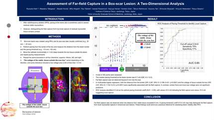
This study used seven porcine models. In vivo radiofrequency ablation (RFA) was performed percutaneously under general anesthesia to create a transmural, box-shaped lesion on the posterior wall of the right atrium. After ablation, pacing threshold measurements were taken from the lesion center using an ablation catheter, advancing in 1-mm increments in four directions (superior, inferior, left, and right). At each point, the pacing threshold (mA) and distance from the center were recorded. If pacing failed at 20 mA, the threshold was recorded as 21 mA.
Following these measurements, the heart was excised for macroscopic confirmation of the lesion’s size and transmurality. The lesion border was defined based on its gross dimensions. The distance from the border was then calculated using the gross lesion size and the distance from the center, and this value was used for statistical analyses. Distances toward the scar (inward from the border) were assigned positive values, and those extending into viable tissue (outward from the border) were assigned negative values. For example, if the lesion measured 20 × 20 mm, the border would lie 10 mm from the center; a point at 7 mm from the lesion center would be +3 mm (inside the scar), whereas a point at 12 mm would be −2 mm (outside).
A total of 392 points were assessed. Even within the same porcine model, the voltage of viable tissue outside the box lesion varied by direction. The median pacing threshold at the lesion border was 6.7 mA (IQR: 4.0–12.0). Far-field capture was not observed beyond 6 mm into the scar. In multivariate logistic regression, both the distance from the border (OR: 0.287, 95% CI: 0.196–0.421, p<0.0001) and the voltage of tissue outside the box (OR: 2.87, 95% CI: 1.742–5.213, p<0.0001) were significantly associated with far-field capture. In contrast, contact force and local scar voltage were not significant predictors. ROC analysis identified 8.9 mA as the optimal pacing threshold cutoff (AUC = 0.75), with values ≥8.9 mA indicating far-field capture and values <8.9 mA suggesting local capture.
Far-field capture was not observed when the distance from viable tissue exceeded 6 mm. A pacing threshold cutoff of 8.9 mA may help distinguish far-field capture from local myocardial capture in transmural scar lesions. These findings could serve as a practical reference for assessing lesion viability after RFA.
Contributors

M Takigawa
Author
M Honda
Author
M Negishi
Author
R Tateishi
Author
I Kawamura
Author
K Yamao
Author
K Goto
Author
T Nishimura
Author
S Tao
Author
K Terata
Author
S Miyazaki
Author
H Watanabe
Author
T Sasano
Author
You may be interested in