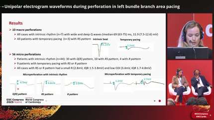
Unipolar electrogram waveforms during perforation in left bundle branch area pacing
European Heart Journal


Abstract
Perforation is common during left bundle branch area pacing (LBBAP) implantation, as the conduction fibers lie just beneath the left ventricular endocardium. Whereas overt (macro) perforation is usually straightforward to diagnose due to loss of capture, recognizing micro perforation, in which only the tip of the lead screw perforates, is much more challenging as thresholds are usually preserved. The main parameters to evaluate perforation are pacing impedance and current of injury (COI) amplitude from the unipolar unfiltered electrogram (EGM). Besides COI amplitude, there are few data on distinct EGM waveforms which are related to perforation.
Our aim was to investigate waveforms of unipolar unfiltered EGM during perforation and compare them to those of the final accepted lead position.
All patients with LBBAP implantation in whom perforation was indicated in our prospective database and which was annotated by a timestamp in the recording system were included in the analysis. Micro perforation was suspected in case of COI <5 mV together with a drop in impedance by >200 Ohms. Macro perforation was defined if in addition there was loss of capture at 5V/0.5ms. Unfiltered (0.5 – 500 Hz) unipolar sensed tip EGM waveform during perforation was compared to that in the final lead position in the same patient. The negative and positive deflections ("Q/S" and "R" waveforms) and COI amplitudes were measured by digital calipers of the recording system.
There were 120/622 cases with at least one perforation, of which 66 (mean age 80 ± 11 years, 62% men) had a time stamp which enabled measurement of the EGM at the time of perforation and at the final position. We recognized 3 distinct patterns of EGMs during perforation: Q(R), RS and R. Most cases (86%) had wide and deep Q/S waves (Table). No Q waves were visible if the sensed beats originated from temporary pacing (n=11, 17%); in those cases, the pattern was RS (n=6) or R (n=5). In the final accepted position, the most common pattern was R with large COI; although 15% patients had a Q wave it was markedly shorter in duration and of lower amplitude than in the perforated beats (Figure).
Ten (15%) patients had macro perforations; the remaining 56 (85%) patients had micro perforations. In those with macro perforation, all cases without temporary pacing showed a wide and deep Q wave (67ms, IQR 66-70ms; 11.9mV, IQR 11.2-12.4mV). In those with micro perforation, 31 had a Q(R) pattern (Q-wave duration 56ms, IQR 50-68mV; 6.0mV, IQR 4.4-10.6mV), 14 had an RS pattern (four with temporary pacing) and eight had an R pattern (four with temporary pacing). The cases with RS or R pattern had a small R (2.8mV, IQR 1.5-3.8mV) and low COI (3.4mV, IQR 1.7-4.8mV).
The unipolar EGM from lead tip showed very distinct patterns during perforations compared to the final position. A deep and wide negative deflection, in addition to low COI amplitude, is indicative of perforation.
Contributors


You may be interested in