Observed differences in right- vs. left-side lesions in the ventricular septum and epicardial vs. endocardial lesions in the free wall, created by bipolar radiofrequency ablation for thick myocardium
European Heart Journal


Abstract
Bipolar (BIP) radiofrequency (RF) ablation may induce differing lesion volume and local temperature rises between the right- and left-side of the interventricular septum (IVS), and between the endocardium and epicardium of the left ventricular (LV) free wall. If so, outcomes of BIP ablation would vary depending on the location of the arrhythmogenic substrate within the myocardial wall.
Experimental study (approved by our Institution) was performed to clarify how lesions created by BIP ablation were different between both sides of either the IVS or LV.
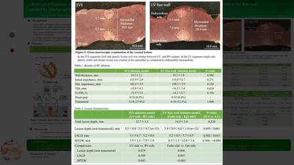
Two inner plastic containers, each almost half the size of the outer experimental container and communicating through windows, were set in the outer experimental container (Figure). Each inner container was filled with Tyrode’s solution at 37 °C, circulated by a pump and temperature-controlled by a heater. Healthy porcine hearts were prepared for coronary perfusing IVS or LV segments, which were placed between the two inner plastic containers. For LV ablation, another plastic board was inserted into one of the inner plastic containers, facing epicardial myocardium, to create a small space to minimize the circulation of Tyrode’s solution and to mimic the pericardial spaces (Figure). Two ablation catheters were placed on opposite surfaces of each myocardium, and BIP ablation (30W, 20-g contact, 120-seconds, 13-ml/min irrigation) was attempted with 18 applications each in IVS and LV segments.
There were no differences in the wall thickness at the ablation sites, initial impedance, total impedance decline (TID) and %TID between IVS and LV ablations. No steam-pop occurred in all applications in IVS and LV ablations. Transmural lesions were created in 5/18 applications in IVS ablation and in 6/18 applications in LV ablation. Total lesion depth (sum of the depth on both sides in non-transmural lesions, and myocardial thickness at the ablation site in the transmural lesions) was not different between IVS and LV ablations (Table). In the non-transmural lesions, epicardial lesion depth was deeper than that in the endocardium in LV ablation but this difference was not observed between RV- and LV-side septum in IVS ablation (Table). Epicardial longest surface lesion diameter (Long-SLD) and maximum lesion width under the surface (Max-LUS) were longer than those in the endocardial lesions in LV ablation (Table), but these parameters were not different between RV- and LV-side septum in IVS ablation.
BIP ablation for IVS created similar lesions in the RV- and LV-side of septum whereas BIP ablation for LV induced larger lesions in epicardial than in endocardial wall, probably due to limited cooling effects in the pericardial space. Physicians need to pay attention for possible excessive temperature rise in the pericardial space during the BIP LV ablation, especially via the temperature monitoring from the ablation catheter placed in the LV cavity.
Contributors

You may be interested in