An improvement in the global longitudinal strain reflecting the mechanical burden of premature ventricular complexes (PVCs) after treatment of PVCs
European Heart Journal


Abstract
We previously reported that the difference in the global longitudinal strain (GLS) between sinus beats (SBs) and SBs immediately after PVCs (SBs after PVCs) indicated the mechanical burden of the PVCs and tension load after an ineffective contraction on the left ventricular (LV) myocardium. An increase in this mechanical burden could contribute to the development of PVC-cardiomyopathy.
This study aimed to investigate the outcomes of treatments to decrease the burden of the PVCs by evaluating the GLS values of SBs, PVCs, and SBs after PVCs and repeating the GLS evaluations after the PVC treatment.
Consecutive patients referred to our institution because of frequent PVCs were recruited for the study and we obtained informed consent. Patients with structural heart disease were excluded. Patients who successfully achieved a full GLS evaluation were investigated. Patients who met all the criteria underwent repeated full GLS evaluations after PVC treatment including medications and catheter ablation.
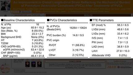
Of 47 patients recruited for the study, 16 (Age: 61±15, Male: 8, LV ejection fraction [LVEF]: 58.3±6.5%, PVC origins: the right ventricular outflow tract [n=11], LV outflow tract [n=3] and others [n=2], number of PVCs: 16264±10624 beats/day) met all criteria and received medications (beta blockers) and/or catheter ablation. Subsequently, patients achieved a 67% reduction in the PVC burden (Figure 1: Baseline GLS evaluation and PVC burden pre- and post-treatment). Repeated echocardiography, including a GLS evaluation after treatment, showed a significant improvement in the SBs-GLS (Figure 2: -16.2±3.7 [pre-] vs. -20.0±4.1 [post-treatment], paired difference: 3.8±2.9, p<0.001), however, the changes in the LVEF, LV diameter (end-diastolic [LVDd] / systolic [LVDs]) did not reach statistical significance (LVEF: 59.4±4.9 vs. 62.1±5.4%, p=0.081, LVDd: 48.3±5.6 vs. 46.9±5.1, p=0.166, LVDs: 32.9±5.9 vs. 30.9±5.4mm, p=0.059. pre- vs. post-treatment, respectively).
Repeated GLS evaluations revealed an improvement in the myocardial fiber contraction, which was not observed by measurements such as the LVEF, after treatment to decrease the PVC burden.
Contributors



You may be interested in