Brain atrophy and cognitive function in atrial fibrillation: a subanalysis of the SKAF study
European Heart Journal


Abstract
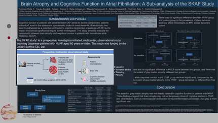
Cognitive function in patients with non-valvular atrial fibrillation (NVAF) tends to decline compared to patients without NVAF, even in the absence of symptomatic stroke or overt dementia. While previous studies have suggested an association between NVAF, and silent cerebral ischemia and microbleeds, the mechanisms linking NVAF to cognitive impairment remain unclear. Brain atrophy has been hypothesized as a potential contributor to cognitive dysfunction in NVAF patients, but its impact and clinical significance require further investigation. This study aimed to evaluate the relationship between brain atrophy and cognitive function in patients with NVAF.
The SKAF study is a prospective, investigator-initiated, multicenter, observational study involving Japanese patients with NVAF aged 60 years or older. We planned to enroll 200 NVAF patients with no history of symptomatic stroke or dementia, including those receiving anticoagulant therapy with edoxaban or not, and 100 patients with sinus rhythm for comparison. Participants underwent brain magnetic resonance imaging (MRI) and cognitive function assessment, including the Montreal Cognitive Assessment (MoCA), at enrollment and after 24 months. We analyzed cerebral atrophy using MRI.
The study enrolled 310 patients (age: 72.1±6.5 years; male: 191 [61.6%]; NVAF: 213 [68.7%]). The mean CHA2DS2-VASc score in the NVAF group was 2.6±1.2. There was no significant difference between the NVAF group and the control group in the prevalence of silent ischemic stroke or silent microbleeds on MRI across the entire cohort. At enrollment, there was no significant difference in the MoCA score between the two groups (NVAF: 24.4±3.4, control: 25.2±2.9, P=0.054), and no difference in the extent of gray matter atrophy (NVAF: 3.5±1.6%, control: 3.4±2.1%, P=0.63). After 24 months, cognitive function in the NVAF group declined significantly compared to the control group (NVAF: 24.2±3.8, control: 25.5±3.2, P<0.01), while the extent of gray matter atrophy in the NVAF group remained no different from that in the control group (NVAF: 3.9±2.7%, control: 3.7±2.0%, P=0.68).
The extent of gray matter atrophy was not directly related to cognitive function in patients with NVAF. These findings suggest that brain atrophy may not be the primary driver of cognitive decline in NVAF, and other factors, such as microvascular dysfunction or neuroinflammatory processes, may play a more significant role.
Contributors


Y Kondo
Author
K Senoo
Author
S Ryuzaki
Author
M Nakano
Author
T Kajiyama
Author
M Nakano
Author
Y Kobayashi
Author
You may be interested in