Atrial fibrillation burden and enlarged perivascular spaces in basal ganglia
European Heart Journal


Abstract
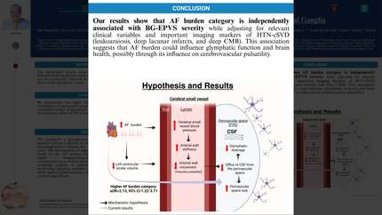
Perivascular spaces (PVS) within the brain are filled with cerebrospinal fluid (CSF) and are important components of the glymphatic system, which is responsible for clearing metabolic waste and maintaining cerebral homeostasis. Vascular pulsatility is a central driver for the propulsion of CSF out from PVS, and when impaired, could lead to MRI-visible PVS enlargement (EPVS) and glymphatic dysfunction. Atrial fibrillation (AF) is a major effector of vascular pulsatility.
We hypothesized that a higher AF burden is associated with the presence of a severe degree of EPVS in the basal ganglia (BG-EPVS), potentially through AF-induced changes in vascular pulsatility.
We analyzed data from 646 consecutive AF patients who had no history of symptomatic stroke, MRI-defined embolic stroke, or history of neurodegenerative disease and who received brain MRIs with appropriate sequences at a tertiary care center. Neuroimaging data were collected by a stroke neurologist blinded to the clinical data. To estimate surrogates of higher and lower levels of AF burden, we classified AF burden into two groups, namely lower (paroxysmal/persistent AF) or higher (longstanding persistent/permanent AF). This classification was validated in a subgroup of 84 patients with quantitative AF burden data (mean AF burden: 86.5% higher vs. 12.9% lower). The imaging outcome variables were the presence of severe BG-EPVS, white matter disease (using Fazekas score), Brain atrophy (using atrophy score), lacunar infarcts, and cerebral microbleeds (CMB) in deep/lobar locations. The multivariable model was adjusted for all variables with p<0.1 in univariate analysis.
In total, 102/646 (15.8%) patients had a severe degree of BG-EPVS, and those patients were older (79.4 ± 8.4 years vs 72.5 ± 9.4 years, p < 0.001), had higher AF burden category (31.4% vs 16.7%, p<0.001), higher CHA2DS2-VASC scores (mean 4.1±1.3 vs 3.5±1.5, p<0.001), and more severe MRI markers of hypertensive cerebral small vessel disease (HTN-cSVD). In multivariable analyses, higher AF burden remained an independent predictor of BG-EPVS (adjusted OR=2.13, 95% CI 1.22-3.71, p=0.008), together with Fazekas score (p<0.001), deep lacunar infarcts (p<0.001), deep CMBs (p=0.041), and age (p<0.001) after adjusting for CHA2DS2-VASc score, serum creatinine, obstructive sleep apnea, and lobar lacunar infarcts and CMBs.
Our results show that AF burden is independently associated with severe BG-EPVS while adjusting for relevant clinical variables and important imaging markers of HTN-cSVD (white matter hyperintensities, deep lacunar infarcts, and deep CMB). This association suggests that AF burden could influence glymphatic function and brain health, possibly through its influence on cerebrovascular pulsatility.
Contributors

S Greenberg
Author
K Helmer
Author
E Gurol
Author
Massachusetts General Hospital - Harvard Medical School Boston , United States of America
O Rotschild
Author
Massachusetts General Hospital - Harvard Medical School Boston , United States of America
B Le
Author
E Gokcal
Author
A Abramovitz Fouks
Author
A Das
Author
You may be interested in