Structural remodeling and left atrial fibrosis in patients with atrial fibrillation and metabolic syndrome
European Heart Journal


Abstract
To compare the area of low voltage zones and parameters characterizing left atrial (LA) dilation in patients with atrial fibrillation (AF) in combination with metabolic syndrome (MS) and without it.
We examined 69 patients with symptomatic paroxysmal AF with ineffective antiarrhythmic therapy. Average age was 55.4±7.7 years. Patients with ischemic heart disease, heart surgery history, chronic heart failure classes II-IV, valvular pathology, systemic and oncological diseases, acute and chronic inflammatory diseases, patients with liver, kidney and thyroid pathology were not included in the study. All patients were divided into two groups: 33 patients with AF combined with MS (n=33) and 36 patients with AF without MS (n=36). Electroanatomic mapping of LA was performed before radiofrequency isolation of pulmonary veins on a sinus rhythm using a ThermoCool SmartTouch catheter with electroanatomical mapping system CARTO3. Using CARTO3 in the off-line mode, the low voltage area of the LA myocardium was estimated. The volume and volume index of the left atrium were determined by echocardiography. Metabolic syndrome was diagnosed in the presence of three or more components according to the criteria of the International Diabetes Federation, 2005.
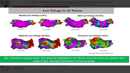
It was established that the area of low LA voltage in patients with AF in combination with MS was greater than in patients with AF without MS (23.2 [13.8;37.0] % and 13.6 [9.9;21.7] %, p=0.0063). A diffuse distribution of low voltage area (anterior and posterior walls of the LA) was more frequent in patients with AF in combination with MS than in patients with AF without MS (66.7% and 33.3%, p=0.01). The volume of LA in patients with AF in combination with MS was greater than in patients with AF without MS (82.0±20.4 and 70.1±20.7, p= 0.0473). There were no significant differences in the LA volume index in the two groups (p= 0.2679). ROC analysis showed that LA volume >73.5 ml in patients with AF will increase the probability of diffuse distribution of low voltage LA zones by 5.7-fold (OR=5.68, 95% CI 1,892-17,099, p=0.002). Meanwhile, LA volume index > 34 ml/m2 will increase the probability of detecting diffuse distribution of low voltage LA zones by 9.3-fold (OR=9.31, 95% CI 2.086-30.686, p=0.002).
Diffuse distribution low voltage area of myocardial left atrial is more frequently detected in patients with paroxysmal atrial fibrillation in combination with metabolic syndrome and is associated with left atrial dilation.
Contributors

You may be interested in