The relationship between calcium thickness and stent expansion by optical coherence tomography for concentric or eccentric calcified lesion after intravascular lithotripsy
European Heart Journal


Abstract
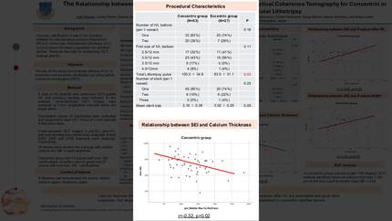
Coronary calcification is one of predictor for suboptimal stent expansion. Shockwave intravascular lithotripsy (IVL) is a useful device that modifies calcified lesions.
We investigated that efficacy of IVL to concentric and eccentric calcifications by optical coherence tomography (OCT). The aim of this study was whether relationship between calcium fractures after IVL and stent expansion by OCT assessment.
A total of 80 patients who underwent OCT-guided IVL and coronary stenting were included. Cross-sectional OCT images at 1-mm intervals in pre-IVL (2,398), post-IVL (2,380), and post-stenting (2,728) time points were analyzed.
All lesions were divided into 2 groups with median calcium arc 180゚in each segments. Concentric group had 53 lesions with over 180゚ calcifications, eccentric calcium group had 27 lesions with less than 180゚ calcifications.
Baseline maximum calcium thickness was similar between 2 groups. Final stent expansion index (SEI) was also similar between 2 groups.
Calcium fractures after IVL were more frequent in concentric group compared with eccentric group (32.0% vs. 23.3%, p=0.03). Stent under-expansion (SEI < 0.85) was associated with maximum calcium thickness in concentric group (p=0.03), but not in eccentric group (p=0.56). In concentric group, receiver operation characteristic curve analysis identified maximum calcium thickness 1,225μm as the best cutoff to predict SEI < 0.85. Percentage of fractured segment after IVL was positively correlated with SEI (r=0.337, p=0.01). Fracture width after IVL was also correlated with SEI (r=0.377, p< 0.01).
Calcium fractures after IVL were more frequent in concentric calcification compared with eccentric calcification. Percentage of fractures and large fracture width after IVL are associated with higher stent SEI. Our study suggests that calcium thickness over 1,225μm is predicted for stent under-expansion in concentric calcified lesions.
Contributors

D Sato
Author
Y Sugiyama
Author
M Kai
Author
H Miyashita
Author
K Shimizu
Author
T Ochiai
Author
N Moriyama
Author
K Tobita
Author
T Matsumoto
Author
S Mizuno
Author
F Yamanaka
Author
Y Tanaka
Author
M Murakami
Author
S Saito
Author
You may be interested in