The impact of lipid rich plaque on coronary-microvascular-dysfunction evaluated by near-infrared-spectroscopy IVUS and angiography-derived index of microcirculatory resistance after PCI
European Heart Journal


Abstract
Coronary microvascular dysfunction (CMD) after percutaneous coronary intervention (PCI) has been reported to be associated not only with periprocedural myocardial infarction, but also with poor prognosis. While index of microcirculatory resistance using pressure wire (invasive IMR) has been conventionally used to evaluate CMD, angio-derived IMR (angio-IMR) has recently emerged as a noninvasive method and has shown good correlation with invasive IMR. Previous investigations have already shown that the maximum value of lipid core burn index (LCBI) for any of the 4-mm segments (maxLCBI4mm) by near-infrared spectroscopy IVUS (NIRS-IVUS) represents the degree of lipid rich plaque (LRP) and can predict microvascular obstruction such as slow flow phenomenon after PCI. However, few studies have examined the relationship between LRP assessed by NIRS-IVUS and the extent of IMR.
The purpose of this study was to clarify the relationship between LRP identified by NIRS-IVUS and CMD assessed by angio-IMR in coronary artery disease (CAD) patients and to determine whether adding lipid area in stent (LAS) as a new index to maxLCBI4mm values can further improve the diagnostic performance of CMD after PCI.
We retrospectively evaluated 147 consecutive patients who underwent PCI using NIRS-IVUS for CAD, excluding STEMI, at our hospital from November 2021 to September 2024. LAS was defined as an index reflecting the total amount of LRP at the stenting site and was calculated using the following formula (LAS = stent diameter x stent length x LCBI in stenting area). MaxLCBI4mm, LAS, and angio-IMR were measured in culprit vessel before and after PCI. CMD was defined as angio-IMR≥25. ROC curve analysis was performed to determine the optimal cut-off values of maxLCBI4mm and LAS for angio-IMR≥25 after PCI.
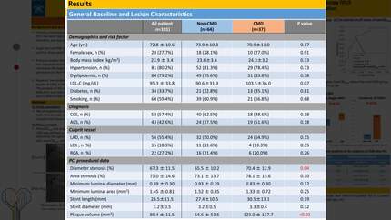
Of 147 patients, CMD after PCI occurred in 54 patients (37%). MaxLCBI4mm and LAS values were significantly higher in the CMD group compared to the non-CMD group (662±199 vs. 422±217, P<0.01; 25984±12967 vs. 13920±10550, P<0.01). Figure 1 shows the results of ROC analysis of maxLCBI4mm and LAS values for predicting the development of CMD after PCI. ROC analysis determined maxLCBI4mm≥579 and LAS≥20384 as an optimal cut-off value associated with development of CMD. Interestingly, patients with higher values of both maxLCBI4mm and LAS were significantly more likely to develop CMD after PCI than those with higher values of either-or lower values of both (Figure 2). On multivariable analysis, maxLCBI4mm≥579 and LAS≥20384 emerged as the independent predictor of the incidence of CMD after PCI (OR=6.9, 95% CI=2.4-21.4, P<0.01; OR=3.1, 95% CI=1.1-8.5, P=0.03).
The results of this study demonstrate that the combination of maxLCBI4mm values and LAS values as a new index in NIRS-IVUS-guided PCI can predict the development of CMD after PCI with high accuracy.
Contributors

You may be interested in